File:Bronchial anatomy.jpg
원본 파일 (2,646 × 2,048 픽셀, 파일 크기: 1.98 MB, MIME 종류: image/jpeg)
캡션
설명
파일 설명[편집]
| 설명 |
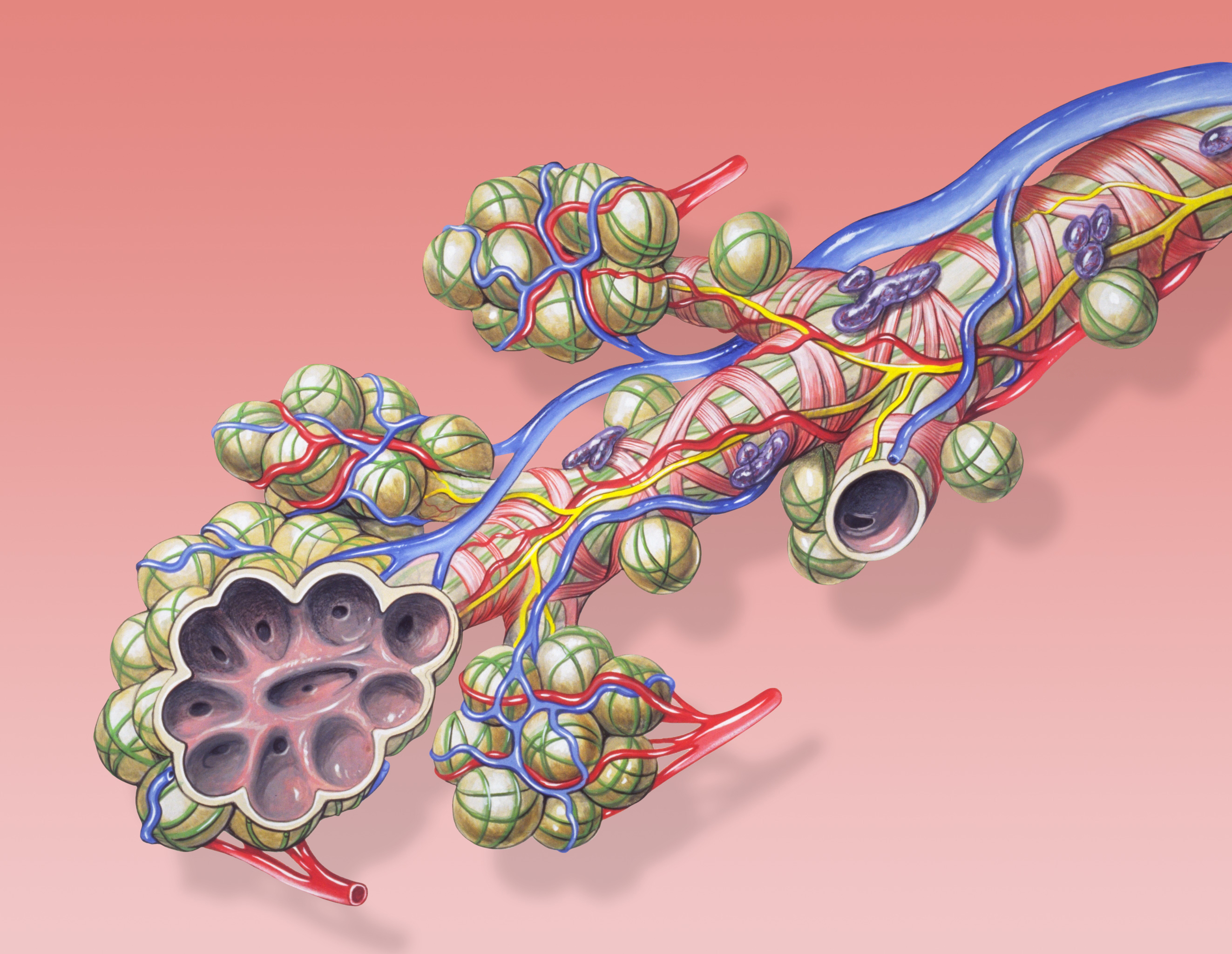
English: Bronchial anatomy detail of alveoli and lung circulation.
Français : Anatomie pulmonaire: détail des alvéoles et de la circulation pulmonaires . |
| 날짜 | |
| 출처 | Patrick J. Lynch, medical illustrator |
| 저자 | Patrick J. Lynch, medical illustrator |
| 저작권 (이 파일을 인용하기) |
Creative Commons Attribution 2.5 License 2006 |
| 다른 버전 | 이 파일은 다음으로 파생됨: Bronchial anatomy Cerchiato.png None |
|

이 이미지는 2012년 1월 16일의 오늘의 이미지로 선정되었습니다. 이미지 설명은 다음과 같습니다. 다른 언어들:
English: Bronchial anatomy detail of alveoli and lung circulation. Español: Anatomía bronquial: detalle de los alvéolos y la circulación pulmonar. Français : Anatomie pulmonaire : détail des alvéoles et de la circulation pulmonaires. Italiano: L'anatomia delle ramificazioni terminali dell'albero respiratorio con l'annessa vascolarizzazione (arteria e vene polmonari). Nederlands: Anatomisch detail van longblaasjes (pulmonaire alveoli) in de longen, waar tijdens de ademhaling de gaswisseling plaatsgrijpt. Русский: Анатомия бронха Українська: Анатомія бронха з розрізом альвеол, бронхіальні частини легеневої артерії і легеневої вени, спинна частина легеневої гілки блукаючого нерва. ქართული: ბრონქების ანატომია დეტალურად. 日本語: 肺胞と肺循環を図解する気管支の解剖図。 中文: 肺泡解刨细节和肺循环。 |
Patrick J. Lynch; illustrator; C. Carl Jaffe; MD; cardiologist Yale University Center for Advanced Instructional Media Medical Illustrations by Patrick Lynch, generated for multimedia teaching projects by the Yale University School of Medicine, Center for Advanced Instructional Media, 1987-2000. Patrick J. Lynch, http://patricklynch.net Creative Commons Attribution 2.5 License 2006; no usage restrictions except please preserve our creative credits: Patrick J. Lynch, medical illustrator; C. Carl Jaffe, MD, cardiologist. https://creativecommons.org/licenses/by/2.5/
라이선스[편집]
- 이용자는 다음의 권리를 갖습니다:
- 공유 및 이용 – 저작물의 복제, 배포, 전시, 공연 및 공중송신
- 재창작 – 저작물의 개작, 수정, 2차적저작물 창작
- 다음과 같은 조건을 따라야 합니다:
- 저작자표시 – 적절한 저작자 표시를 제공하고, 라이센스에 대한 링크를 제공하고, 변경사항이 있는지를 표시해야 합니다. 당신은 합리적인 방식으로 표시할 수 있지만, 어떤 방식으로든 사용권 허가자가 당신 또는 당신의 사용을 지지하는 방식으로 표시할 수 없습니다.
| 주석 | 이 이미지에는 주석이 있습니다: 공용에서 주석을 보기 |
파일 역사
날짜/시간 링크를 클릭하면 해당 시간의 파일을 볼 수 있습니다.
| 날짜/시간 | 섬네일 | 크기 | 사용자 | 설명 | |
|---|---|---|---|---|---|
| 현재 | 2010년 8월 4일 (수) 11:31 | | 2,646 × 2,048 (1.98 MB) | Dcoetzee (토론 | 기여) | Remove watermark |
| 2006년 12월 26일 (화) 04:49 |  | 2,646 × 2,048 (1.42 MB) | Patrick.lynch (토론 | 기여) | {{Information |Description = Bronchial anatomy detail of alveoli and lung circulation |Source = Patrick J. Lynch, medical illustrator |Date = December 23, 2006 |Author = Patrick J. Lynch, medical illustrator |Permission = Creative Commons Attribution 2.5 |
이 파일을 덮어쓸 수 없습니다.
이 파일을 사용하는 문서
다음 문서 40개가 이 파일을 사용하고 있습니다:
- User:Miya/POTD
- User:Ö/Best/2010
- User talk:99of9/Promotions
- Commons:Featured picture candidates/File:Bronchial anatomy.jpg
- Commons:Featured picture candidates/Log/August 2010
- Commons:Featured pictures/Non-photographic media/Computer-generated
- Commons:Featured pictures/chronological/2010-B
- Commons:Picture of the Year/2010/Galleries/Diagrams
- Commons:Picture of the Year/2010/Galleries/Diagrams/Large
- Commons:Picture of the Year/2010/Galleries/Diagrams/Small
- Commons:Picture of the Year/2010/Galleries/Index/9
- Commons:Picture of the Year/2010/Galleries/Index/Diagrams
- Commons:Picture of the Year/2010/Galleries/Table
- Commons:Picture of the Year/2010/Galleries/Table/08
- Commons:Picture of the Year/2010/R1/File:Bronchial anatomy.jpg
- Commons:Picture of the Year/2010/Results/R1/ALL/Table
- Commons:Picture of the Year/2010/Results/R1/Category winners
- Commons:Picture of the Year/2010/Results/R1/Checking
- Commons:Picture of the Year/2010/Results/R1/Diagrams
- Commons:Picture of the Year/2010/Results/R1/Diagrams/Table
- Commons talk:Picture of the Year/2010/Galleries/Table
- Commons talk:Picture of the Year/2010/Results/R1/ALL/Table
- File:Bronchial anatomy Cerchiato.png
- Template:Potd/2012-01
- Template:Potd/2012-01-16
- Template:Potd/2012-01-16 (da)
- Template:Potd/2012-01-16 (de)
- Template:Potd/2012-01-16 (en)
- Template:Potd/2012-01-16 (es)
- Template:Potd/2012-01-16 (fr)
- Template:Potd/2012-01-16 (it)
- Template:Potd/2012-01-16 (ja)
- Template:Potd/2012-01-16 (ka)
- Template:Potd/2012-01-16 (ko)
- Template:Potd/2012-01-16 (mk)
- Template:Potd/2012-01-16 (nl)
- Template:Potd/2012-01-16 (ru)
- Template:Potd/2012-01-16 (uk)
- Template:Potd/2012-01-16 (zh-hans)
- Template:Potd/2012-01 (zh-hans)
이 파일을 사용하고 있는 모든 위키의 문서 목록
다음 위키에서 이 파일을 사용하고 있습니다:
- als.wikipedia.org에서 이 파일을 사용하고 있는 문서 목록
- ar.wikipedia.org에서 이 파일을 사용하고 있는 문서 목록
- az.wikipedia.org에서 이 파일을 사용하고 있는 문서 목록
- ba.wikipedia.org에서 이 파일을 사용하고 있는 문서 목록
- be-tarask.wikipedia.org에서 이 파일을 사용하고 있는 문서 목록
- be.wikipedia.org에서 이 파일을 사용하고 있는 문서 목록
- bg.wikipedia.org에서 이 파일을 사용하고 있는 문서 목록
- bn.wikipedia.org에서 이 파일을 사용하고 있는 문서 목록
- bs.wikipedia.org에서 이 파일을 사용하고 있는 문서 목록
- ckb.wikipedia.org에서 이 파일을 사용하고 있는 문서 목록
- crh.wikipedia.org에서 이 파일을 사용하고 있는 문서 목록
- cs.wikipedia.org에서 이 파일을 사용하고 있는 문서 목록
- cv.wikipedia.org에서 이 파일을 사용하고 있는 문서 목록
- da.wikipedia.org에서 이 파일을 사용하고 있는 문서 목록
- de.wikipedia.org에서 이 파일을 사용하고 있는 문서 목록
- de.wikibooks.org에서 이 파일을 사용하고 있는 문서 목록
- en.wikipedia.org에서 이 파일을 사용하고 있는 문서 목록
- en.wikibooks.org에서 이 파일을 사용하고 있는 문서 목록
- en.wikiversity.org에서 이 파일을 사용하고 있는 문서 목록
- eo.wikipedia.org에서 이 파일을 사용하고 있는 문서 목록
- es.wikibooks.org에서 이 파일을 사용하고 있는 문서 목록
- eu.wikipedia.org에서 이 파일을 사용하고 있는 문서 목록
- fa.wikipedia.org에서 이 파일을 사용하고 있는 문서 목록
- fi.wikipedia.org에서 이 파일을 사용하고 있는 문서 목록
- fr.wikipedia.org에서 이 파일을 사용하고 있는 문서 목록
- gl.wikipedia.org에서 이 파일을 사용하고 있는 문서 목록
이 파일의 더 많은 사용 내역을 봅니다.