Category:Bacteria
Jump to navigation
Jump to search
domain of micro-organisms .jpg) E. coli, 10 000× | |||||||
| Upload media | |||||||
| Instance of | |||||||
|---|---|---|---|---|---|---|---|
| Subclass of | |||||||
| Start time | |||||||
| Different from | |||||||
| |||||||
| |||||||
| Taxon author | Carl Woese, 2024 | ||||||
| |||||||
- Included phyla (for NCBI):
- Acidobacteria, Actinobacteria, Aquificae, Armatimonadetes, Bacteroidetes, Caldiserica, Chlamydiae, Chlorobi, Chloroflexi, Chrysiogenetes, Cyanobacteria, Deferribacteres, Deinococcus-Thermus, Dictyoglomi, Elusimicrobia, Fibrobacteres, Firmicutes, Fusobacteria, Gemmatimonadetes, Lentisphaerae, Nitrospirae, Planctomycetes, Proteobacteria, Spirochaetes, Synergistetes, Tenericutes, Thermodesulfobacteria, Thermotogae, Verrucomicrobia
- Acidobacteria, Actinobacteria, Aquificae, Bacteria incertae sedis, Bacteroidetes, Caldiserica, Chlamydiae, Chlorobi, Chloroflexi, Cyanobacteria, Deferribacteres, Deinococcus-Thermus, Elusimicrobia, Fibrobacteres, Firmicutes, Fusobacteria, Gemmatimonadetes, Lentisphaerae, Nitrospirae, Planctomycetes, Proteobacteria, Spirochaetes, Synergistetes, Tenericutes, Thermodesulfobacteria, Thermotogae, Verrucomicrobia
- Note: Only marine phyla
- Abditibacteriota, Acidobacteriota, Actinomycetota, Aquificota, Armatimonadota, Atribacterota, Bacillota, Bacteroidota, Balneolota, Caldisericota, Calditrichota, Chlamydiota, Chlorobiota, Chloroflexota, Chrysiogenota, Coprothermobacterota, Cyanobacteriota, Deferribacterota, Deinococcota, Dictyoglomerota, Elusimicrobiota, Fibrobacterota, Fusobacteriota, Gemmatimonadota, Kiritimatiellota, Lentisphaerota, Mycoplasmatota, Nitrospinota, Nitrospirota, Planctomycetota, Pseudomonadota, Rhodothermota, Spirochaetota, Synergistota, Thermodesulfobacteriota, Thermomicrobiota, Thermotogota, Verrucomicrobiota
- Note: Only valid phyla
Subcategories
This category has the following 35 subcategories, out of 35 total.
- Bacteria in Auckland Museum (18 F)
-
.
?
A
- Acidobacteriota (4 F)
B
C
D
- Dictyoglomi (3 F)
F
G
M
- Methylomirabilota (11 F)
N
P
R
S
T
- Thermodesulfobacteriota (6 F)
V
Media in category "Bacteria"
The following 200 files are in this category, out of 249 total.
(previous page) (next page)-
Jer-Bactéthie.ogg 1.0 s; 12 KB
-
De-Bakterie2.ogg 1.8 s; 18 KB
-
De-Bakterium2.ogg 2.0 s; 19 KB
-
 12864 2008 Article 1790 Fig1 HTML.png 1,184 × 486; 314 KB
-
 13068 2022 2210 Fig2 HTML.webp 1,960 × 1,438; 203 KB
-
31. Микробиолошки квалитет на млеко.ogg 4 min 17 s, 1,920 × 1,080; 297.83 MB
-
 96pinner.jpg 2,810 × 2,691; 2.01 MB
-
 A thousands eyes (9404711386).jpg 4,928 × 3,264; 2.47 MB
-
 Agar Art with Microbes.jpg 4,000 × 3,000; 2.27 MB
-
 Agar Plates stored in the laminar flow cabinet.jpg 3,264 × 1,836; 3.02 MB
-
 Amor por bactérias.jpg 1,269 × 1,133; 464 KB
-
 Anaerobic Chamber.pdf 1,650 × 1,275; 4.08 MB
-

-
 Antimicrobial resistance.jpg 6,720 × 4,480; 5.74 MB
-
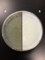
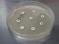 Antimicrobial susceptibility testing (AST) result of Leuconostoc.jpg 4,000 × 3,000; 1.43 MB
-
 Atlas of bacterial flagellation (1960) (19721564194).jpg 1,824 × 2,828; 1.04 MB
-
 Bacteri attachment.jpg 718 × 540; 69 KB
-
 Bacteria (35323217002).jpg 1,149 × 1,564; 243 KB
-
 Bacteria (35795301244).jpg 1,149 × 1,834; 290 KB
-
 Bacteria and pus cells in a Gram stained smear of sputum.jpg 4,000 × 3,000; 1.49 MB
-
 Bacteria beginning to colonize.jpg 1,600 × 1,200; 99 KB
-
 Bacteria Bidirectional DNA Replication.png 1,082 × 903; 417 KB
-
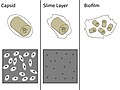 Bacteria Capsules and Slime Layers.jpg 720 × 540; 52 KB
-
 Bacteria collage.jpg 2,880 × 2,160; 3.31 MB
-
 Bacteria growth on everyday objects.png 750 × 1,334; 2.42 MB
-
 Bacteria ico.png 64 × 64; 7 KB
-
 Bacteria in saline wet mount.jpg 4,000 × 3,000; 1.34 MB
-
 Bacteria infect tissues using ‘chemical harpoons’ (17978494003).jpg 1,100 × 790; 937 KB
-
 Bacteria involved in causing and treating cancers.svg 512 × 293; 403 KB
-
 Bacteria on everyday objects.png 750 × 1,334; 1.88 MB
-
 Bacteria Spirillum 6.jpg 1,280 × 1,024; 119 KB
-
 Bacteria Spirillum 7.jpg 1,280 × 1,024; 115 KB
-
 Bacteria Spirillum 8.jpg 1,280 × 1,024; 115 KB
-
 Bacteria Spirillum 9.jpg 1,280 × 1,024; 134 KB
-
 Bacteria template.png 40 × 50; 3 KB
-
 Bacteria through microscope.jpg 2,448 × 3,264; 1.17 MB
-
 Bacteria USP response.png 406 × 311; 20 KB
-
 Bacterial cell walls.jpg 1,012 × 717; 795 KB
-
 Bacterial Cluster (After image enhancement).png 2,048 × 2,048; 2.35 MB
-
 Bacterial Cluster (Before image enhancement).png 2,048 × 2,048; 1.56 MB
-
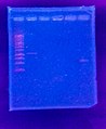 Bacterial DNA in the gel under the UV light.jpg 1,744 × 2,125; 2.28 MB
-
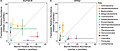
 Bacterial OTUs from clone libraries and next-generation sequencing.png 1,748 × 1,212; 1.3 MB
-
 Bacterial small rna mechanisms.png 1,280 × 720; 110 KB
-
 Bacterial twitching.png 1,143 × 653; 204 KB
-
 Bacterial UTI picture of urine microscopy showing plenty of pus cells and bacteria.jpg 3,264 × 2,448; 1.07 MB
-
 Bacteriapolis at the Exploratorium.jpg 4,504 × 2,772; 5.4 MB
-
 Bacterias lacticas.jpg 1,284 × 762; 73 KB
-
 Bacterias vistas en microscopio electrónico de barrido.jpg 3,072 × 2,082; 7.99 MB
-
 Bactérie acétique.jpg 1,280 × 853; 414 KB
-
 Bakterien (Bacteria) - 630x (14045255442).jpg 3,180 × 2,172; 3.02 MB
-
 Batteri.jpg 640 × 480; 22 KB
-
 BD Bactec for rapid detction of microbial growth.jpg 4,000 × 1,844; 3.94 MB
-
 Bifunctional ppGpp synthase hydrolase SpoT.png 802 × 920; 382 KB
-
 Big Bacteria (43435845060).jpg 4,752 × 3,168; 1.11 MB
-
 Big Bacteria.jpg 800 × 600; 53 KB
-
 Binding of SmeT to DNA.png 603 × 539; 166 KB
-
 Blood culture bottle without growth of organisms.jpg 4,000 × 3,000; 6.03 MB
-
 The relation of bacteria to the flavors of cheddar cheese (IA bullbai062).pdf 814 × 1,300, 37 pages; 3.39 MB
-
 Casein hydrolysis.png 1,856 × 2,475; 4.85 MB
-
 CEM&fungus.jpg 872 × 640; 81 KB
-

-
 Changes in swimmer velocity and Reynolds number with length scale.jpg 1,084 × 516; 291 KB
-
 Chemosynthetic Microbial Mats.jpg 3,000 × 2,250; 1.46 MB
-
 Chocolate agar.jpg 1,844 × 4,000; 3.89 MB
-
 Chromatium2.jpg 750 × 304; 83 KB
-

-
 ColE1 replication control.png 4,068 × 5,324; 572 KB
-
 Colibacile pwels.jpg 234 × 256; 7 KB
-
 COLONIE BATTERICHE ISOLATE NEL TERRENO.jpg 3,870 × 2,591; 5.57 MB
-
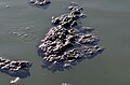
 Colonie batteriche o vita marina?.jpg 2,505 × 2,505; 3.67 MB
-
 Colonization of potato tubers by bacteria.png 1,200 × 1,652; 1.36 MB
-
 ColorazioneCapsula.JPG 198 × 97; 2 KB
-
 Common Bacterial Culture Media.jpg 4,000 × 3,000; 6.39 MB
-
 Compound image from "Functional Green-Tuned Proteorhodopsin from Modern Stromatolites".png 2,150 × 1,637; 5.11 MB
-
 Conditions multiplication thermiques bacteries.png 600 × 822; 34 KB
-
 Connecting.jpg 290 × 190; 13 KB
-
 Control de Plagas.jpg 6,016 × 4,000; 14.67 MB
-
-
 Culture Plates.jpg 4,752 × 3,168; 6.26 MB
-
 Cyanase enzymatic reaction scheme.png 1,018 × 270; 39 KB
-
 Cyanase Pentamer with labeled argenine resides.png 1,554 × 1,482; 554 KB
-
 Dark water running from a pool.jpg 3,672 × 4,896; 9.16 MB
-
 Diagnostic algorithm of possible bacterial infection.png 5,376 × 4,133; 2.77 MB
-
 Die Gartenlaube (1879) b 063 1.jpg 1,027 × 865; 569 KB
-
 Die Gartenlaube (1891) b 362 2.jpg 252 × 301; 8 KB
-
 Die Gartenlaube (1891) b 363 1.jpg 390 × 248; 12 KB
-
 Die Gartenlaube (1891) b 363 5.jpg 275 × 279; 8 KB
-
 Die Gartenlaube (1891) b 364.jpg 118 × 326; 4 KB
-
 Die Pflanzenwelt (1913-1922.) (20913876076).jpg 2,038 × 3,218; 1.2 MB
-
 Divergence from a common ancestor.png 360 × 320; 91 KB
-
 DNA barcoding of marine bacteria.jpg 500 × 129; 90 KB
-
 DNA Extraction of Bacteria.jpg 4,000 × 3,000; 6.42 MB
-
 DNA Replication in prokaryotes.png 2,000 × 729; 408 KB
-
 Dust storms as a source of aerosolized bacteria.png 460 × 239; 122 KB
-
 E.coli harverd medical school.jpg 1,280 × 720; 53 KB
-
 Effect of penicillin on the coli bacteria.jpg 1,522 × 1,831; 2.94 MB
-
 Effector binding to SmeT.png 557 × 538; 313 KB
-
 EnteroPluri Test.png 3,852 × 458; 470 KB
-
 Essential metabolic genes in bacteria.png 720 × 540; 79 KB
-
 Extreme aquatic habitats and their extremophiles and molecules.png 12,430 × 6,581; 5.26 MB
-
 Extrinsic Drug Resistance.jpg 4,000 × 3,000; 6.57 MB
-
 Fermentación de azúcares.jpg 3,896 × 3,240; 2.18 MB
-
 Fight Bac! (085 086) (7396068296).jpg 7,200 × 5,400; 6.97 MB
-
 Figure2b.pdf 1,125 × 450; 73 KB
-
 Figure2d.pdf 1,125 × 450; 100 KB
-
 Figure2e.pdf 1,125 × 450; 112 KB
-
 Flagellar Motor Assembly.jpg 3,840 × 2,160; 1.12 MB
-
 FLoc vase Basse-Deûle rive G vers St André61.JPG 4,928 × 3,264; 1.27 MB
-
 Fmicb-11-602250-g001D.jpg 549 × 879; 118 KB
-
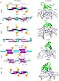 Fold Topology and Oligomerization States of H-NS.jpg 663 × 919; 210 KB
-
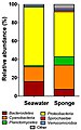
 Freshwater versus marine bacterial abundance by taxonomy.jpg 1,280 × 549; 81 KB
-
 Gas formation by bacteria in TSI agar tube.jpg 3,264 × 2,448; 1.59 MB
-
 GenericBacteriumClipArt.pdf 141 × 54; 2 KB
-
 Glucosa-lactosa-sacarosa.jpg 3,896 × 3,240; 2.19 MB
-
 Gram Negative Rods in Sputum Gram Staining.jpg 4,000 × 2,250; 1 MB
-
 Gram Positive Cocci.jpg 4,000 × 2,250; 1.23 MB
-
 Gram's collor.jpg 3,264 × 1,836; 475 KB
-
 Grand Prismatic Spring Bacteria PLC-PK-YS-15.jpg 3,661 × 1,457; 4.61 MB
-
 Green running water.jpg 3,672 × 4,896; 9.09 MB
-
 Greenish bacteria and concretions.jpg 4,896 × 3,672; 8.78 MB
-
 Growth of bacteria on MHA isolated from sewage.jpg 3,000 × 4,000; 3.42 MB
-
 Growth of microbes in blood culture bottle.jpg 4,000 × 3,000; 3.71 MB
-
 Größen von Partikeln in Aerosolen.png 1,748 × 1,128; 67 KB
-
 Host killing.png 5,391 × 3,568; 424 KB
-
 Host parasite interaction.jpg 4,000 × 2,250; 1.15 MB
-
 Immune Response to Exotoxins.png 1,500 × 1,125; 239 KB
-
 Indole Test Positive.jpg 4,000 × 3,000; 2.86 MB
-
 Interaction of bacteria with variously shaped cargoes.jpg 963 × 384; 222 KB
-
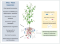
 Introducing plant beneficial bacteria into plant seed 2.jpg 2,025 × 891; 139 KB
-
 Introducing plant beneficial bacteria into plant seed.jpg 2,159 × 1,559; 228 KB
-
 Iron Bacteria in Bankhead National Forest.JPG 1,600 × 1,137; 1.09 MB
-
 Koloni morfolojisi.png 678 × 600; 26 KB
-
 Lab WT.jpg 2,448 × 3,264; 1.41 MB
-
 Laboratory bacterial culture.jpg 3,024 × 4,032; 2.24 MB
-
 Lactose fermenting colonies of Escherichia coli.jpg 4,000 × 3,000; 1.34 MB
-
 Leuconostoc antibiogram result.jpg 4,000 × 3,000; 1.42 MB
-
 Leuconostoc mesenteroides Gram Staining.jpg 4,000 × 3,000; 1.32 MB
-
 Leuconostoc mesenteroides growth on CLED agar.jpg 4,000 × 3,000; 1.46 MB
-
 Leuconostoc mesenteroides in wet mount.jpg 4,000 × 2,250; 1.62 MB
-
 Light-induced bacterial infiltration into a leaf.jpg 641 × 766; 140 KB
-
 Lip print in bacteria on agar plate.jpg 3,264 × 2,448; 2.4 MB
-
 LPS-gl.svg 433 × 1,054; 85 KB
-
 Luminous bacterial species in light organ symbiosis in fish and squid.png 1,000 × 3,826; 1.17 MB
-
 Made for one instant (31375179818).jpg 4,752 × 3,168; 1.45 MB
-
 Magnetic Beads Spider.tif 3,072 × 2,045; 5.57 MB
-
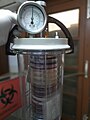 McIntosh and Fildes’ Anaerobic Jar.jpg 3,000 × 4,000; 5.89 MB
-
 Meat diet bacteria.jpg 1,949 × 991; 275 KB
-
 MED152 Transporters.png 3,000 × 1,400; 263 KB
-

-
 Metabolic reconstruction of a member of the Delphibacteria phylum.png 1,810 × 1,088; 699 KB
-
 Meyers b2 s0275a.jpg 1,478 × 2,353; 2.38 MB
-
 MIC estimation agar dilution technique.jpg 2,904 × 2,592; 2.94 MB
-
 Microbial Art.jpg 4,000 × 3,000; 2.55 MB
-
 Microorganisms-10-01169-g006.png 3,606 × 1,768; 1.77 MB
-
 Micropia.jpg 5,146 × 3,430; 12.75 MB
-
 Miscroscope slide with bacteria.JPG 640 × 480; 136 KB
-
 Mitose simple lv.jpg 446 × 619; 46 KB
-
 Model bacterium with a spheroidal cell body and a helical flagellum.png 1,085 × 218; 19 KB
-

-
 Mold?.jpg 5,184 × 3,456; 9.48 MB
-
 Morphology and ultrastructure of Ca. T. magnifica.jpg 1,454 × 2,006; 302 KB
-
 Motility cartoon type 1a.jpg 300 × 300; 17 KB
-
 Motility cartoon type 1b.jpg 350 × 250; 29 KB
-
 Motility cartoon type 1c.jpg 350 × 250; 19 KB
-
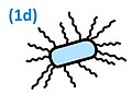 Motility cartoon type 1d.jpg 350 × 250; 21 KB
-
 Motility cartoon type 8b.jpg 300 × 300; 26 KB
-
 Mucoid non-lactose fermenting E. coli.jpg 4,000 × 3,000; 1.39 MB
-
 Musty wine.jpg 4,288 × 2,848; 2.27 MB
-
 Negative Staining Examples (8530733273).jpg 968 × 1,128; 509 KB
-
 Non-lactose fermenting colonies of E. coli on CLED agar.jpg 4,000 × 3,000; 1.13 MB
-
 Numerous Gram Negative Bacteria and Pus cells in Gram staining of sputum.jpg 4,000 × 2,250; 1,020 KB
-
 Orange running water.jpg 3,672 × 4,896; 8.95 MB
-
 Oxidase test positive bacteria and AST.jpg 4,000 × 3,000; 5.06 MB
-
 Oxidase Test Positive.jpg 4,000 × 3,000; 1.58 MB
-
 Oxygen requirement of bacteria.png 757 × 579; 489 KB
-
 Packed pus cells and bacteria in urine sediment microscopy.jpg 3,264 × 2,448; 1.24 MB
-
 Pairs of marine and freshwater samples sharing common taxa.jpg 1,280 × 637; 95 KB
-
 Patchclamp Spheroplast1.jpg 3,072 × 2,304; 1.45 MB
-

-
 PBR322.jpg 460 × 440; 40 KB
-
 Peptidoglycan L-Lys-D-Glu.png 1,352 × 1,182; 19 KB
-
 Plant responses to bacteria in the rhizosphere.webp 3,077 × 2,240; 1.26 MB
-
 Plant-endophytic bacteria interactions.webp 2,500 × 3,499; 1.59 MB
-

-
 Pone.0023479.g001.png 2,092 × 4,432; 2.19 MB
-
 Porcellio scaber (10.3897-zookeys.801.22395) Figure 7.jpg 1,512 × 751; 646 KB
-
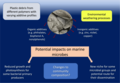 Potential impacts of plastic pollution on marine bacteria.webp 1,899 × 1,313; 147 KB
-

-
 Prokaryote-Tree-41467 2019 12171 Fig1.webp 1,740 × 1,465; 277 KB
-
 Prosthecochloris aestuarii culture.jpg 2,604 × 4,624; 1.79 MB
-
 Prosthecochloris aestuarii.png 511 × 391; 158 KB
-
 Purple bacteria.jpg 530 × 398; 86 KB
-
 Pus cells, Epithelial cells, RBCs and Bacteria in Urine Microscopy.jpg 4,000 × 2,250; 2.1 MB
-
 R. Muir, Bacteriological Atlas, 1927 Wellcome L0030995.jpg 3,386 × 3,340; 4.04 MB
-
 R. Muir, Bacteriological Atlas, 1927 Wellcome L0030996.jpg 1,458 × 1,447; 820 KB
-
 R. Muir, Bacteriological Atlas, 1927 Wellcome L0030997.jpg 3,360 × 3,376; 3.99 MB
-
 R. Muir, Bacteriological Atlas, 1927 Wellcome L0030998.jpg 1,440 × 1,416; 791 KB
-

-
-
 Rapid plasma reagin (RPR) Test Reactive for syphilis.jpg 3,264 × 2,448; 2.37 MB


.jpg)


_result_of_Leuconostoc.jpg)
_(19721564194).jpg)

.jpg)
.jpg)


.jpg)


.png)
.png)


_-_630x_(14045255442).jpg)


.jpg)


_b_063_1.jpg)
_b_362_2.jpg)
_b_363_1.jpg)
_b_363_5.jpg)
_(20913876076).jpg)


_(7396068296).jpg)


.jpg)


.jpg)


.jpg)


_(14580380047).jpg)


_Figure_7.jpg)


_Test_Reactive_and_Non-reactive_Results_Demonstration_for_Syphilis.jpg)
_Test_Reactive_for_syphilis.jpg)
.jpg)
_b_364.jpg)