Category:Cell nucleus
Перейти до навігації
Перейти до пошуку
membrane-bounded organelle of eukaryotic cells in which chromosomes are housed and replicated | |||||
| Завантажити медіафайл | |||||
| Вимова (аудіофайл) | |||||
|---|---|---|---|---|---|
| Є одним із |
| ||||
| Є підкласом |
| ||||
| Частина від | |||||
| Фізично взаємодіє з | |||||
| Не плутати з | |||||
| |||||
Підкатегорії
Показано 11 підкатегорій із 11.
- SVG cell nucleus (28 F)
A
C
- Cell nucleus shape (8 F)
D
- Cell nucleus division (22 F)
H
N
- Nuclear transfer techniques (2 F)
P
Файли в категорії «Cell nucleus»
Показано 200 файлів цієї категорії (із 357).
(попередня сторінка) (наступна сторінка)-
De-Zellkern.ogg 2,0с; 19 КБ
-
 202012 Nuclear membrane.svg 512 × 512; 657 КБ
-
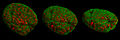 3D-SIM-2 Nucleus prophase 3d rotated.jpg 1920 × 640; 377 КБ
-
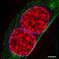 3D-SIM-3 Prophase 3 color.jpg 902 × 896; 386 КБ
-
Actin-nucleation-at-the-centrosome-controls-lymphocyte-polarity-ncomms10969-s2.ogv 3,3с, 501×250; 128 КБ
-
-
-
-
-
-
 BarrBodyBMC Biology2-21-Fig1clip293px.jpg 293 × 148; 28 КБ
-
-
-
 Blausen 0212 CellNucleus ru.png 1600 × 1785; 2,15 МБ
-
 Blausen 0212 CellNucleus.png 1600 × 1785; 2,34 МБ
-
 Bovine Pulmonary Artery Endothelial Cells Fluorescent Image.jpg 1920 × 1452; 304 КБ
-
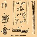
 Brockhaus-Efron Yadro kletki 1.jpg 552 × 862; 129 КБ
-
 Brockhaus-Efron Yadro kletki 2.jpg 761 × 752; 93 КБ
-
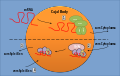
 Cajal bodies.jpg 414 × 378; 57 КБ
-
 Cajal-Body-Detail.svg 1134 × 720; 253 КБ
-
 Cajal-Body-Overview.svg 1134 × 720; 132 КБ
-
 Cell nucleus 1 -- Smart-Servier.png 2350 × 2502; 971 КБ
-
 Cell nucleus 2 -- Smart-Servier.png 3400 × 1488; 456 КБ
-
 Cell nucleus 3 -- Smart-Servier.png 1484 × 1640; 284 КБ
-
 Cell nucleus I -- Smart-Servier.jpg 10 240 × 5760; 1,84 МБ
-
 Cell nucleus II -- Smart-Servier.jpg 10 240 × 5760; 1,4 МБ
-
-
-
-
-
-
 Chromosome cohesion - en.png 960 × 720; 26 КБ
-
 Chromosome cohesion.png 652 × 482; 11 КБ
-
 Cicle de la Ran-GTP.PNG 2000 × 1350; 688 КБ
-
-
-
-
-
-
-
-
-
-
-
-
-
-
-
De-Nukleus.ogg 2,0с; 19 КБ
-
 Diagram human cell nucleus no text.png 310 × 296; 41 КБ
-
 Diagram human cell nucleus serbian nuclear envelope.PNG 462 × 378; 46 КБ
-
 Diagram human cell nucleus serbian nuclear pore.PNG 462 × 378; 46 КБ
-
 Diagram human cell nucleus serbian nucleolus.PNG 462 × 378; 46 КБ
-
 Diagram human cell nucleus serbian nucleoplasm.PNG 462 × 378; 46 КБ
-
 Diagram human cell nucleus sk.jpg 462 × 378; 76 КБ
-
 Diagram human cell nucleus.png 462 × 378; 49 КБ
-
 Diagram showing where genes are in cells CRUK 380.png 396 × 396; 63 КБ
-
-
-
-
 DNA recycle hypothes.PNG 600 × 563; 259 КБ
-
 Draq5-fireRGB-bar.tif 1024 × 1024; 3 МБ
-
-
-
-
-
-
 Esquema de nucli cel·lular.PNG 552 × 346; 45 КБ
-
-
-
-
-
-
-
Fast-imaging-of-live-organisms-with-sculpted-light-sheets-srep09385-s1.ogv 4,1с, 960×540; 4,04 МБ
-
Fast-imaging-of-live-organisms-with-sculpted-light-sheets-srep09385-s2.ogv 4,1с, 960×540; 2,61 МБ
-
Fast-imaging-of-live-organisms-with-sculpted-light-sheets-srep09385-s3.ogv 4,1с, 960×540; 2,93 МБ
-
 FcRn-mediated IgG Recycling.png 528 × 393; 49 КБ
-
 Flemming1882Tafel1Fig14.jpg 458 × 469; 75 КБ
-
-
-
-
-
-
 GFP Superresolution Christoph Cremer.JPG 538 × 389; 156 КБ
-
 HeLa pspecks2.jpg 283 × 298; 46 КБ
-
-
-
-
-
-
-
-
-
-
-
-
-
-
 Human Tpp1.png 300 × 300; 30 КБ
-

-
-
-
-
-
-
-
-
-
-
-
-
 Interno del nucleo.png 366 × 230; 150 КБ
-
 Intranuclear structures EM.jpg 1604 × 946; 417 КБ
-
-
-
-
-
-
-
 Kernmatrix.png 670 × 423; 7 КБ
-
 Kinetochore vertebrates-en.png 1920 × 720; 73 КБ
-
 Kinetochore vertebrates.png 1370 × 538; 27 КБ
-
 Kodola uzbūve.svg 473 × 349; 108 КБ
-
-
-
-
 Leeuwenhoek1719RedBloodCells.jpg 1440 × 489; 54 КБ
-
Light-Sheet-Microscopy-for-Single-Molecule-Tracking-in-Living-Tissue-pone.0011639.s004.ogv 6,7с, 256×128; 4,15 МБ
-
Light-Sheet-Microscopy-for-Single-Molecule-Tracking-in-Living-Tissue-pone.0011639.s005.ogv 6,7с, 384×128; 1,51 МБ
-
Light-Sheet-Microscopy-for-Single-Molecule-Tracking-in-Living-Tissue-pone.0011639.s006.ogv 6,7с, 128×128; 2,13 МБ
-
Light-Sheet-Microscopy-for-Single-Molecule-Tracking-in-Living-Tissue-pone.0011639.s007.ogv 6,7с, 128×128; 1,44 МБ
-
-
-
-
-
-
-
-
-
-
MDCK-Cystogenesis-Driven-by-Cell-Stabilization-within-Computational-Analogues-pcbi.1002030.s019.ogv 1хв 20с, 550×550; 1,46 МБ
-
MDCK-Cystogenesis-Driven-by-Cell-Stabilization-within-Computational-Analogues-pcbi.1002030.s020.ogv 1хв 20с, 550×550; 1,54 МБ
-
-
-
-
-
-
-
Mechanical-interplay-between-invadopodia-and-the-nucleus-in-cultured-cancer-cells-srep09466-s2.ogv 1хв 0с, 976×800; 9,51 МБ
-
-
Mechanical-interplay-between-invadopodia-and-the-nucleus-in-cultured-cancer-cells-srep09466-s4.ogv 1хв 0с, 992×784; 17,78 МБ
-
-
-
-
-
-
-
-
-
-
-
 Microtubule.png 793 × 771; 1,11 МБ
-
-
 Nuclear components ru.jpg 459 × 152; 30 КБ
-
 Nuclear envelope of one cancerous HeLa cell.png 2553 × 1577; 1,6 МБ
-
 Nuclear grooves.jpg 196 × 163; 14 КБ
-
 Nuclear membrane.png 720 × 311; 109 КБ
-
-
-
-
-
-
-
-
 NuclearSpeckle-Splicing.svg 832 × 720; 587 КБ
-
-
-
-
-
 Nucleus ER.png 416 × 469; 31 КБ
-
 Nucleus Nucleolus and chromatin of animal cell.png 349 × 348; 245 КБ
-
 Nucleus of a cell (diagram).jpg 815 × 688; 247 КБ
-
 Nucleus of cardiac fibroblasts.jpg 3456 × 4608; 4,44 МБ
-
 Nucleus.png 300 × 286; 42 КБ
-
 Nucléole.JPG 441 × 353; 22 КБ
-
 Numt insertion.jpg 358 × 274; 26 КБ
-
-
-
-
-
-
-
 O.Hertwig1906Fig2-6.jpg 1382 × 1459; 347 КБ
-
Optogenetic-control-of-nuclear-protein-export-ncomms10624-s3.ogv 12с, 457×433; 193 КБ
-
-
-
-
-


.jpg)


