Category:Cellulose
Перайсьці да навігацыі
Перайсьці да пошуку
палімэрнае злучэньне   | |||||
| Загрузіць мэдыя | |||||
| Асобны выпадак панятку |
| ||||
|---|---|---|---|---|---|
| Падкляса ад |
| ||||
| Частка ад |
| ||||
| Названа ў гонар | |||||
| Першаадкрывальнік | |||||
| Прызначэньне |
| ||||
| Складаецца з | |||||
| Маса (вага) |
| ||||
| Ня блытаць з | |||||
| |||||
Падкатэгорыі
Гэтая катэгорыя зьмяшчае наступныя 12 падкатэгорыяў з 12 агулам.
B
- Bacterial cellulose (10 F)
C
- Cellophane (17 F)
- Cellulose insulation (20 F)
F
- Cellulose fillers (2 F)
M
- Mother of vinegar (10 F)
P
V
- Vinegar syndrome (16 F)
Файлы ў катэгорыі «Cellulose»
Паказаныя 119 файлаў гэтай катэгорыі з 119.
-
De-Cellulose.ogg 1,6 с; 17 кб
-
De-Zellulose.ogg 1,7 с; 16 кб
-
 1 Delepierre Gwendoline Cellulose Nanocrystal Fingerprints.jpg 2468 × 3096; 7,61 Мб
-
 1 Delepierre Gwendoline VanGogh Cellulose Nanocrystals.jpg 4140 × 3096; 7,54 Мб
-
 11 Zellulosevorhang 1350x1800.jpg 1350 × 1800; 522 кб
-
 219 Three Important Polysaccharides-01.jpg 1582 × 547; 357 кб
-
 Alkylketendimere Reaktion mit Cellulose.svg 542 × 122; 39 кб
-
 ASA-Reaktion mit Cellulose.svg 426 × 400; 99 кб
-
 Attisholz01 cropped.jpg 869 × 550; 137 кб
-
 Attisholz01.jpg 2640 × 1760; 3,77 Мб
-
 Attisholz02.jpg 1760 × 2640; 4,2 Мб
-
 Car-CNC.jpg 840 × 952; 131 кб
-
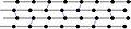 Catene cellulosa.jpg 371 × 84; 9 кб
-
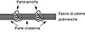 Catene cellulosiche.jpg 294 × 112; 7 кб
-
 Cellulase 1JS4.jpg 507 × 245; 56 кб
-
 Cellulos to glycose.png 1275 × 252; 129 кб
-
 Cellulosa.svg 744 × 1052; 28 кб
-
 Cellulosa1.jpg 240 × 210; 37 кб
-
 Cellulose Acetate disposable syringe filter.jpg 4000 × 3000; 2,1 Мб
-
 Cellulose degradation by certain microorganism.jpg 4608 × 3456; 3,09 Мб
-
 Cellulose formula & Chemical Pulping.tiff 1250 × 1078; 199 кб
-
 Cellulose Haworth.svg 245 × 179; 14 кб
-
 Cellulose II between.gif 490 × 372; 25 кб
-
 Cellulose membranes applying the phase separation method.jpg 1414 × 694; 120 кб
-
 Cellulose nanocrystals.jpg 300 × 300; 89 кб
-
 Cellulose nanofiber network.jpg 963 × 746; 111 кб
-
 Cellulose Nanotechnology (36902405632).jpg 768 × 769; 148 кб
-
 Cellulose Nanotechnology (36902406032).jpg 805 × 1125; 650 кб
-
 Cellulose Nanotechnology (36931269271).jpg 447 × 291; 120 кб
-
 Cellulose polymer.jpg 1024 × 600; 88 кб
-
 Cellulose Sessel.svg 385 × 178; 20 кб
-
 Cellulose spacefilling model.jpg 1070 × 419; 121 кб
-
 Cellulose strand-es.jpg 422 × 372; 115 кб
-
 Cellulose strand.svg 402 × 364; 75 кб
-
 Cellulose Structural Formula V1.svg 619 × 246; 10 кб
-
 Cellulose structure.jpg 869 × 467; 68 кб
-
 Cellulose structure.png 3723 × 2304; 1,26 Мб
-
 Cellulose-2D-skeletal.png 1100 × 488; 44 кб
-
 Cellulose-2D-skeletal.svg 1061 × 570; 3 кб
-
 Cellulose-3D-balls.png 1000 × 570; 211 кб
-
 Cellulose-3D-vdW.png 1000 × 610; 150 кб
-
 Cellulose-Ibeta-from-xtal-2002-3D-balls.png 2000 × 748; 379 кб
-
 Cellulose-Ibeta-from-xtal-2002-3D-vdW.png 2000 × 863; 511 кб
-
 Cellulose-Ibeta-from-xtal-2002-CM-3D-balls.png 2000 × 753; 419 кб
-
 Cellulose-Pyrolyse zu LGO.svg 312 × 416; 108 кб
-
 Cellulose.jpg 3508 × 2480; 196 кб
-
 Cellulose.png 3920 × 103; 3 кб
-
 Cellulose2.PNG 1001 × 398; 26 кб
-
 Cellulosebiochem.png 452 × 166; 9 кб
-
 CelluloseRXN.png 1085 × 1254; 31 кб
-
 Cellulosomes1.jpg 1920 × 2560; 1,44 Мб
-
 Celulose.png 525 × 121; 5 кб
-
 Celuloza-es.jpg 390 × 328; 40 кб
-
 Celuloza.jpg 390 × 328; 25 кб
-
 Chitin glucose and cellulose.svg 512 × 274; 290 кб
-
 Comparison of different cellulose-based membranes.jpg 675 × 779; 81 кб
-
 Crossed Polar Graffiti.tif 1200 × 900; 2,36 Мб
-
 DEAE cellulose preparation.svg 815 × 177; 24 кб
-
 EB1911 Cellulose-lignone.jpg 69 × 61; 3 кб
-

-
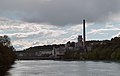
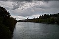
 Ehemalige Cellulosefabrik an der Aare, Attisholz SO 20211009-jag9889.jpg 4608 × 2880; 7,6 Мб
-
 Eskema zelulosa.svg 497 × 288; 43 кб
-
 Estructura celulosa.png 674 × 113; 2 кб
-
 Estrutura da Celulose.png 605 × 113; 4 кб
-
 Fibre-carta.jpg 1600 × 1200; 758 кб
-
 Fibreboard 07877.jpg 3162 × 2372; 2,55 Мб
-
 Foreign body embolism - IVDA (4337751538).jpg 1280 × 960; 506 кб
-
 Foreign body embolization in an intravenous drug abuser - Case 256 (8515416475).jpg 2272 × 1704; 2,75 Мб
-
 Foreign body embolization in an intravenous drug abuser - Case 256 (8515416833).jpg 2272 × 1704; 3,29 Мб
-
 Foreign body embolization in an intravenous drug abuser - Case 256 (8516530054).jpg 2272 × 1704; 3,45 Мб
-

-
 HaworthStrukturCellulose.jpg 3369 × 797; 152 кб
-
 Hemicellulose acting in the plant cell wall.jpg 3988 × 2151; 1,16 Мб
-
 Homopolysaccharide.svg 315 × 259; 22 кб
-
 Illustration of a lignocellulosic structure.jpg 675 × 433; 64 кб
-
 Illustration of the cellulose composition.jpg 712 × 724; 57 кб
-
 Intramolecular glucose carbon Isotopes from tree rings.png 526 × 540; 51 кб
-
 Liaisons hydrogène entre molécules de cellulose.png 405 × 261; 3 кб
-
 Liaisons hydrogène entre molécules de cellulose.svg 462 × 230; 83 кб
-
 Lyocell.jpg 1657 × 1813; 385 кб
-

-
 Mecanismos de reacción de la celulosa.png 562 × 104; 2 кб
-
 Microfibrilles de cellulose légendé.png 723 × 329; 16 кб
-
 Musée de la viscose (Échirolles) abc29.JPG 2288 × 1712; 911 кб
-
 Nanocrystalline-cellulose-I--without-water-molecules.jpg 600 × 270; 67 кб
-
Native-cellulose-nanofibrills-induce-immune-tolerance-in-vitro-by-acting-on-dendritic-cells-srep31618-s2.ogv 12 с, 1440 × 1080; 2,64 Мб
-
-
Native-cellulose-nanofibrills-induce-immune-tolerance-in-vitro-by-acting-on-dendritic-cells-srep31618-s4.ogv 12 с, 1440 × 1080; 3,39 Мб
-
-
 OrganoClick Technology.gif 1071 × 391; 8 кб
-
 Paper. Magnification power 2000x. 01.jpg 6000 × 8000; 5,28 Мб
-
 Paper. Magnification power 2000x. 02.jpg 6000 × 8000; 5,29 Мб
-
 Plant cell showing primary and secondary wall by CarolineDahl.jpg 3015 × 2236; 878 кб
-
 Polymère cellulose.PNG 2257 × 744; 101 кб
-
 Preparation of cellulose-VPBA mixed solution.jpg 1904 × 1616; 370 кб
-
 Preparation of the modified cellulose-based complex particle.jpg 610 × 496; 86 кб
-
 Pumping the insulation in.jpg 1600 × 1060; 322 кб
-
 Rayon closeup 1.jpg 300 × 200; 28 кб
-
 RGB cellulose pigments and glitter - Droguet Benjamin, University of Cambridge.jpg 6016 × 4000; 3,02 Мб
-
 Russellllinters.jpg 560 × 417; 147 кб
-
 S08-06-celljuloza.jpg 306 × 56; 5 кб
-
 Sample Isotherm Fig 1a.jpg 448 × 330; 13 кб
-
 Sample Isotherm Fig 1B.jpg 448 × 330; 11 кб
-
 Saurer Celluloseabbau.svg 194 × 260; 57 кб
-
-
 Schematics of microbial mechanisms of lignocellulose degradation.jpg 670 × 966; 147 кб
-
 Self assembeled-cellulose nanocrystals.jpg 640 × 480; 63 кб
-
 Structure de la paroi pectocellulosique.svg 1052 × 744; 98 кб
-
 Structure of a bacterial cellulose synthase.png 929 × 785; 308 кб
-
 Strukturformel Zellulose.png 674 × 113; 2 кб
-
 Tapetenkleister.jpg 500 × 470; 43 кб
-
 TEMPO-oxidation of cellulose.jpg 1315 × 786; 64 кб
-
 VSReactiveGroups Reaction V2.svg 375 × 70; 38 кб
-
 VSReactiveGroups Reaction.svg 672 × 129; 61 кб
-
 Xanthation.png 2620 × 705; 130 кб
-
 XylanWoodsubunit.png 2241 × 644; 98 кб
-
 Zellstoffverbundelemente.JPG 1632 × 1232; 772 кб
-

-
 Workman operating a guncotton press behind protective rope screen.tiff 3251 × 2086; 25,87 Мб


.jpg)
.jpg)
.jpg)


_(20997061828).jpg)


.jpg)
.jpg)
.jpg)
.jpg)
.jpg)


..jpg)

_abc29.JPG)


