Category:Human brain (horizontal section)
Vai alla navigazione
Vai alla ricerca

anatomical plane that divides the body into superior and inferior parts | |||||
| Carica un file multimediale | |||||
| Sottoclasse di |
| ||||
|---|---|---|---|---|---|
| |||||
Sottocategorie
Questa categoria contiene un'unica sottocategoria, indicata di seguito.
File nella categoria "Human brain (horizontal section)"
Questa categoria contiene 52 file, indicati di seguito, su un totale di 52.
-
 3DTomo.jpg 409 × 544; 59 KB
-
 Axial basal-ganglia.jpg 487 × 441; 64 KB
-
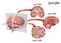 Blausen 0104 Brain x-secs SectionalPlanes-ar.jpg 2 250 × 1 600; 463 KB
-
 Blausen 0104 Brain x-secs SectionalPlanes-Arabic-YM.png 2 250 × 1 600; 2,21 MB
-
 Blausen 0104 Brain x-secs SectionalPlanes.png 2 250 × 1 600; 1,85 MB
-
 Brain; transverse section of cerebral hemisphere. Wellcome L0002346.jpg 1 150 × 1 624; 590 KB
-
 CT-scan-4th-and-3rd-ventricles.JPG 730 × 708; 71 KB
-
 Encefalo.png 849 × 1 026; 1,17 MB
-
 Face sup T1.png 563 × 482; 222 KB
-
 Fahr syndrome.gif 914 × 473; 83 KB
-
 Functional magnetic resonance imaging.jpg 250 × 208; 11 KB
-
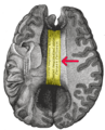 Gray 733-emphasizing-corpus-callosum.png 405 × 500; 793 KB
-
 Gray 737-emphasizing-corpus-callosum.png 500 × 589; 465 KB
-
 Gray716.png 500 × 523; 81 KB
-
 Gray717-emphasizing-external capsule.png 500 × 568; 194 KB
-
 Gray730.png 500 × 383; 29 KB
-
 Gray733.png 405 × 500; 58 KB
-
 Gray737.png 500 × 589; 105 KB
-
 Gray740.png 266 × 494; 52 KB
-
 Gray742-emphasizing-claustrum.png 500 × 572; 193 KB
-
 Gray742.png 500 × 572; 57 KB
-
 Gray746.png 329 × 650; 21 KB
-
 Gray748.png 500 × 510; 66 KB
-
 Gray750.png 417 × 516; 79 KB
-
 Head Plastination - Transverse Section.jpg 2 448 × 2 920; 1 023 KB
-
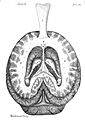
 Head revealing skull, Vesling "Syntagma", 1647 Wellcome L0007899.jpg 1 166 × 1 622; 766 KB
-
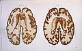
 Horizontal sections of fetal brain.jpg 960 × 720; 123 KB
-
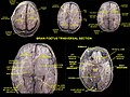
 J. M. Charcot, Diseases of the nervous syste Wellcome L0029909.jpg 4 140 × 2 586; 4,46 MB
-
 Kasraie scalped 3d010r cal BET2 0001.png 405 × 405; 61 KB
-
 Laika ac Semmelweis Museum (10441856403).jpg 3 232 × 4 935; 8,49 MB
-
 Mesencephalon1.jpg 812 × 531; 115 KB
-
 Mts ca mammae T2.jpg 396 × 428; 28 KB
-
 Mésencéphale22.jpg 812 × 531; 116 KB
-
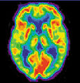 NIH PET.JPG 692 × 721; 294 KB
-
 NPH MRI 274.gif 512 × 512; 2,79 MB
-
 PET3.jpg 300 × 368; 15 KB
-
 Pick's disease.png 1 200 × 552; 223 KB
-
 Principles of Psychology (James) v1 p38.png 1 530 × 2 130; 878 KB
-
 Putamen.jpg 1 181 × 1 423; 324 KB
-
 Pvx8133.tmp.jpg 339 × 297; 18 KB
-
 Regeczy780.jpg 1 308 × 1 167; 158 KB
-
 Schizophrenia PET scan.jpg 224 × 248; 23 KB
-
 Slide2GRE.JPG 960 × 720; 83 KB
-
 Slide3GRE.JPG 960 × 720; 79 KB
-
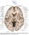
 Sobo 1909 635.png 1 045 × 1 043; 3,12 MB
-
 Sobo 1909 637.png 1 061 × 1 050; 3,19 MB
-
 T1map brain.png 256 × 256; 59 KB
-
 Telencephalon-Horiconatal.jpg 500 × 572; 78 KB
-
 User-FastFission-brain.gif 213 × 231; 444 KB
-
 VBM3.jpg 303 × 393; 15 KB
-
 Vesalius 609c.png 923 × 736; 400 KB
-
 Visible Human head slice.jpg 468 × 590; 70 KB


.jpg)


_v1_p38.png)


