Category:Mikael Häggström/Micrographs of the urinary bladder
Jump to navigation
Jump to search

These images were created by Mikael Häggström, M.D.
- User info
- Reusing images
Media in category "Mikael Häggström/Micrographs of the urinary bladder"
The following 14 files are in this category, out of 14 total.
-
 Cytology of normal urothelial cells, Pap stain.jpg 1,609 × 981; 257 KB
-
 Cytopathology of atypical urothelial cells (AUC).jpg 1,569 × 1,277; 303 KB
-
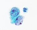
 Cytopathology of high-grade urothelial carcinoma (HGUC).jpg 2,048 × 1,833; 609 KB
-
 Cytopathology of reactive urothelial changes (original).jpg 2,048 × 1,532; 247 KB
-
 Cytopathology of reactive urothelial changes.png 2,385 × 1,429; 3.28 MB
-
 Cytopathology suspicious for high-grade urothelial carcinoma (suspicious HGUC).jpg 1,017 × 1,005; 191 KB
-
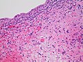
 Histopathology of high grade urothelial carcinoma.jpg 2,048 × 1,333; 728 KB
-
 Histopathology of radiation cystitis.jpg 2,048 × 1,532; 708 KB
-
 Histopathology of squamous cell carcinoma of the urinary bladder, high magnification.jpg 2,048 × 1,532; 433 KB
-
 Histopathology of squamous cell carcinoma of the urinary bladder, low magnification.jpg 2,048 × 1,532; 855 KB
-
 Histopathology of urothelial carcinoma invading the detrusor muscle.jpg 2,048 × 1,532; 940 KB
-
 The Paris System for reporting urinary cytology 2.0.png 5,265 × 4,537; 7.59 MB
-
 The Paris System for reporting urinary cytology.png 5,376 × 3,800; 6.66 MB
-
 Urine cytology with red blood cells.jpg 1,546 × 1,155; 256 KB

.jpg)
.jpg)
.jpg)

.jpg)


