Category:Mikael Häggström/X-rays of the lower extremity
Jump to navigation
Jump to search

This category includes the hip joint.
These images were created by Mikael Häggström, M.D.
- User info
- Reusing images
Media in category "Mikael Häggström/X-rays of the lower extremity"
The following 53 files are in this category, out of 53 total.
-
 Danis–Weber classification on X-ray.jpg 1,108 × 1,267; 290 KB
-
 Danis–Weber classification on X-ray.svg 554 × 633; 182 KB
-
 Intraoperative acetabular fracture, annotated.jpg 602 × 617; 85 KB
-
 Intraoperative acetabular fracture, original.jpg 1,053 × 2,137; 372 KB
-
 Knee prosthesis zones by Knee Society 2015, AP view.jpg 1,809 × 1,453; 334 KB
-
 Knee prosthesis zones by Knee Society 2015, AP view.svg 905 × 726; 152 KB
-
 Knee prosthesis zones by Knee Society 2015, lateral view.jpg 1,176 × 2,198; 383 KB
-
 Knee prosthesis zones by Knee Society 2015, lateral view.svg 588 × 1,099; 192 KB
-
 Lateral and medial joint space of patella.jpg 1,091 × 554; 89 KB
-
 Lateral and medial joint space of patella.svg 546 × 277; 150 KB
-
 Lateral patellofemoral angle.jpg 1,121 × 551; 87 KB
-
 Lateral patellofemoral angle.svg 561 × 276; 149 KB
-
 Metatarsal pseudo-epiphysis.jpg 916 × 1,164; 232 KB
-
 Metatarsal pseudo-epiphysis.svg 458 × 582; 241 KB
-
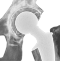
 Postoperative radiograph of hip prosthesis - anteroposterior view (transparent).png 1,033 × 2,096; 580 KB
-
-
 Postoperative radiograph of hip prosthesis - anteroposterior view.jpg 1,033 × 2,096; 396 KB
-
 Postoperative radiograph of hip prosthesis - lateral view.jpg 907 × 2,165; 325 KB
-
 Postoperative radiograph of hip prosthesis - lateral view.png 907 × 2,165; 963 KB
-
 Postoperative X-ray of normal knee prosthesis, anteroposterior view, annotated.jpg 1,918 × 4,156; 753 KB
-

-
 Postoperative X-ray of normal knee prosthesis, anteroposterior view.jpg 959 × 2,078; 249 KB
-
 Postoperative X-ray of normal knee prosthesis, lateral view, annotated.jpg 2,164 × 4,200; 824 KB
-
 Postoperative X-ray of normal knee prosthesis, lateral view, annotated.svg 1,082 × 2,100; 186 KB
-
 Postoperative X-ray of normal knee prosthesis, lateral view.jpg 1,082 × 2,100; 310 KB
-
 Skin folds close to a hip fracture (with arrows).jpg 575 × 587; 107 KB
-
 Skin folds close to a hip fracture.jpg 1,127 × 761; 260 KB
-
 Skin folds over a hip fracture.jpg 1,509 × 895; 269 KB
-
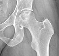
 X-ray of a normal hip.jpg 1,263 × 1,071; 415 KB
-
 X-ray of a normal knee by anteroposterior projection.jpg 1,067 × 1,913; 244 KB
-
 X-ray of a normal knee by lateral projection.jpg 1,133 × 1,939; 286 KB
-
 X-ray of a normal patella.jpg 684 × 433; 34 KB
-
 X-ray of Hansson pins for mildly compressed hip fracture.jpg 1,318 × 1,066; 260 KB
-
 X-ray of Hansson pins with contact points.jpg 1,247 × 747; 195 KB
-
 X-ray of Hansson pins.jpg 1,247 × 747; 191 KB
-
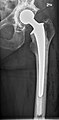
 X-ray of hip with total arthroplasty - Anteroposterior.jpg 1,168 × 2,083; 472 KB
-
 X-ray of hip with total arthroplasty - Lateral.jpg 1,013 × 1,711; 354 KB
-
 X-ray of idiopathic avascular necrosis of the femoral head - Anteroposterior.jpg 1,151 × 1,085; 425 KB
-
 X-ray of idiopathic avascular necrosis of the femoral head - Lateral.jpg 983 × 1,087; 287 KB
-
 X-ray of mildly compressed hip fracture, annotated.jpg 1,755 × 1,047; 334 KB
-
 X-ray of mildly compressed hip fracture.jpg 1,755 × 1,047; 332 KB
-
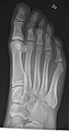
 X-ray of normal right foot by dorsoplantar projection.jpg 1,061 × 2,431; 817 KB
-
 X-ray of normal right foot by lateral projection.jpg 2,638 × 1,322; 662 KB
-
 X-ray of normal right foot by oblique projection.jpg 1,077 × 2,029; 558 KB
-
 X-ray of osteogenesis imperfecta type 5 in newborn - left leg.jpg 433 × 1,595; 139 KB
-
 X-ray of osteogenesis imperfecta type 5 in newborn - right leg - annotated.jpg 467 × 1,559; 128 KB
-
 X-ray of osteogenesis imperfecta type 5 in newborn - right leg.jpg 467 × 1,559; 127 KB
-
 X-ray of pelvis with idiopathic avascular necrosis of the femoral head.jpg 1,283 × 1,051; 309 KB
-
 X-ray of pelvis with total arthroplasty.jpg 2,591 × 2,155; 1.04 MB
-
 X-ray of subtle compressive hip fracture, labeled.jpg 815 × 879; 175 KB
-
 X-ray of subtle compressive hip fracture.jpg 815 × 879; 173 KB
-
 X-ray of the pelvis of a 22 months old male - anteroposterior.jpg 1,271 × 1,023; 286 KB
-
 X-ray of the pelvis of a 22 months old male - Lauenstein.jpg 1,467 × 943; 261 KB


.jpg)


.png)