Category:Mitosis
Vai alla navigazione
Vai alla ricerca
metodo di riproduzione cellulare  | |||||
| Carica un file multimediale | |||||
| Istanza di | |||||
|---|---|---|---|---|---|
| Sottoclasse di | |||||
| Consiste di |
| ||||
| Distinto da | |||||
| |||||
Sottocategorie
Questa categoria contiene le 20 sottocategorie indicate di seguito, su un totale di 20.
A
C
- Cyclin B1 (7 F)
- Cyclin-dependent kinase 1 (11 F)
D
- Mitotic cells stained with DAPI (25 F)
M
- Mitotic signaling (9 F)
P
- Preprophase band (9 F)
- Prometaphase (8 F)
S
T
Pagine nella categoria "Mitosis"
Questa categoria contiene un'unica pagina, indicata di seguito.
File nella categoria "Mitosis"
Questa categoria contiene 154 file, indicati di seguito, su un totale di 154.
-
 0300 Flourescence Stained.jpg 675 × 645; 225 KB
-
 5 Makhluk Mitologi yang diyakini Dunia.pdf 1 500 × 843, 7 pagine; 1,88 MB
-
-
 Anaphase during Mitosis.svg 512 × 394; 32 KB
-
 Attivazione MPF (semplice).png 1 067 × 449; 15 KB
-
 Cell cycle (5 stages of mitotic cell life).jpg 2 200 × 500; 457 KB
-
 Cell division according to A. Schneider (1873).png 1 262 × 357; 83 KB
-
 Cell division according to E. Russov (1872).png 1 090 × 787; 98 KB
-
 Cell division according to I. D. Chistyakov (1874).png 520 × 569; 52 KB
-
 Cell division according to W. Flemming (1882).png 1 286 × 1 652; 414 KB
-
 Cell division process.jpg 766 × 556; 30 KB
-
 Cell polarity.jpg 181 × 160; 7 KB
-
 Cell proliferation.jpg 1 557 × 876; 96 KB
-
 Cervical AIS, ThinPrep.jpg 634 × 643; 212 KB
-
 Chromatin bridge stained using DAPI 1.tiff 1 392 × 1 040; 4,14 MB
-
 Chromosomes in mitosis and meiosis.png 396 × 373; 17 KB
-
 Chromosomes2.jpg 256 × 256; 41 KB
-
 Cleavage Furrow Regression.svg 536 × 461; 11 KB
-
 Cleavage Furrow Regression.tif 572 × 492; 19 KB
-
 Com mROS contribueix a la senyalització mitogènica..gif 441 × 522; 787 KB
-
 Complete network.jpg 1 391 × 867; 98 KB
-

-
 CONDENSING CHROMOSOMES 2.jpg 1 935 × 549; 785 KB
-
 Cormophyta alternancia-de-generaciones.png 192 × 192; 9 KB
-
 Critique of the Theory of Evolution Fig 046.jpg 510 × 820; 94 KB
-
 Cuerpo Flemming Asimetrica.jpg 680 × 900; 92 KB
-
 Cuerpo Flemming Simetrico.jpg 692 × 900; 95 KB
-
 Cuerpo Flemming Ubicacion.png 592 × 434; 276 KB
-
 Cuerpo intermedio Flemming.png 1 912 × 560; 433 KB
-
 Cytokinesis.png 280 × 302; 4 KB
-
 Diagramatic illustration of the successive stages of mitosis (22699945268).jpg 1 890 × 2 385; 1,29 MB
-
 Difference between Chromosome and Chromatid.png 1 528 × 850; 51 KB
-
 Difference between chromosomes and chromatids.png 1 514 × 838; 110 KB
-
 Diploid Cell.png 1 424 × 792; 121 KB
-
 Division cellule.jpg 400 × 147; 69 KB
-
 DNA Replication in Diploid Cell.png 1 425 × 799; 125 KB
-
 EB1911 Cytology - heterotypical mitosis.jpg 760 × 277; 62 KB
-
 EB1911 Cytology - maturation divisions (1).jpg 813 × 655; 171 KB
-
 EB1911 Cytology - nuclear division.jpg 766 × 935; 274 KB
-
 EB1911 Cytology - preparation for mitosis (2).jpg 891 × 421; 99 KB
-
 EB1911 Cytology - preparation for mitosis.jpg 793 × 211; 52 KB
-
 EB1911 Rhizopoda - Bud-fission of Euglypha alveolata.jpg 1 086 × 738; 307 KB
-
 Embryo in flower.png 3 000 × 3 006; 2,97 MB
-
 Figure 04.jpg 700 × 955; 104 KB
-
 Figure 05.jpg 800 × 605; 65 KB
-
 Figure 06.jpg 400 × 1 025; 56 KB
-
 Figure 10 02 04.jpg 544 × 544; 173 KB
-

-
 Fluxograma - atuação da pRb.jpg 896 × 873; 79 KB
-
 Friedrich Reinke's medical school graduation document.jpg 1 575 × 1 959; 331 KB
-
 Gray2.png 376 × 600; 19 KB
-
 Gyhhhh.jpg 788 × 389; 32 KB
-

-
 Interphase cycle- Mitosis and Meiosis.png 1 416 × 817; 284 KB
-
 Interphase mitosis.png 508 × 149; 2 KB
-
 Irreversible Bistable Switch in Mitotic Exit.jpg 960 × 633; 54 KB
-
 MajorEventsInMitosis.jpg 460 × 167; 19 KB
-
 Malignant spindle cell neoplasm showing mitotic figures 40X.jpg 997 × 749; 287 KB
-
 Malignant spindle cell neoplasm showing mitotic figures.jpg 997 × 749; 273 KB
-
 Meiosis 1- Anaphase 1.png 995 × 728; 77 KB
-
 Meiosis 1- Cytokinesis.png 1 661 × 803; 110 KB
-
 Meiosis 1- Prophase 1.png 998 × 713; 63 KB
-
 Meiosis 1- Telophase 1.png 629 × 733; 50 KB
-
 Meiosis 2- Anaphase 2.png 1 372 × 725; 102 KB
-
 Meiosis 2- Cytokinesis (2).png 1 322 × 777; 92 KB
-
 Meiosis 2- Cytokinesis.png 1 322 × 758; 83 KB
-
 Meiosis 2- Metaphase 2.png 1 370 × 726; 115 KB
-
 Meiosis 2- Prophase 2.png 1 661 × 725; 67 KB
-
 Meiosis 2- Telophase 2.png 1 372 × 725; 95 KB
-
 Meiosis- Metaphase 1.png 630 × 727; 64 KB
-
 Meiosis1- Cytokinesis.png 1 372 × 775; 81 KB
-
 Metaphase during Mitosis.svg 512 × 431; 49 KB
-
 Mitoos.jpg 1 022 × 372; 58 KB
-
 Mitoosi1.jpg 97 × 55; 3 KB
-
 Mitoosi2.jpg 105 × 70; 4 KB
-
 Mitoosi3.jpg 230 × 75; 5 KB
-
 Mitos delar kromosomerna i en cellkärna..png 1 023 × 372; 127 KB
-
 Mitose colchicine fr.svg 800 × 263; 135 KB
-
 Mitose.gif 359 × 501; 4 KB
-
 Mitose.JPG 691 × 600; 48 KB
-
 Mitosiaren faseak - eu.svg 2 361 × 409; 1,16 MB
-
 Mitosis (1).jpg 1 800 × 2 255; 929 KB
-
 Mitosis (13083175463).jpg 595 × 842; 61 KB
-
 Mitosis Animation.gif 1 200 × 675; 324 KB
-
 Mitosis by Elspeth.jpg 1 440 × 1 175; 272 KB
-
 MITOSIS cells secuencie-es.jpg 1 000 × 149; 49 KB
-
 Mitosis cells sequence English.svg 774 × 115; 494 KB
-
 Mitosis classification.png 683 × 734; 136 KB
-
 Mitosis cycle.jpg 2 464 × 2 056; 2,87 MB
-
 Mitosis In A Lymphoma Cell.jpg 715 × 667; 281 KB
-
 Mitosis Mesenchymal Stem Cells.gif 300 × 253; 2,94 MB
-
 Mitosis of onion cells.jpg 3 024 × 4 032; 2,4 MB
-
 Mitosis process.gif 600 × 400; 120 KB
-
 Mitosis schematic diagram-es.svg 833 × 723; 190 KB
-
 Mitosis Stages.JPG 2 592 × 1 936; 2,21 MB
-
 Mitosis stages.jpg 1 564 × 1 564; 1,23 MB
-
 Mitosis vs Meiosis Daughter Cells.png 1 254 × 653; 156 KB
-
 Mitosis- Anaphase.png 625 × 751; 54 KB
-
 Mitosis- Cytokinesis (1).png 1 369 × 663; 84 KB
-
 Mitosis- Cytokinesis.png 1 372 × 756; 69 KB
-
 Mitosis- Metaphase.png 646 × 758; 71 KB
-
 Mitosis- Prophase.png 1 491 × 831; 162 KB
-
 Mitosis- Telophase.png 625 × 753; 36 KB
-
 Mitosis-AscarisEggcs400x2.jpg 1 024 × 768; 140 KB
-
 Mitosis.jpg 158 × 142; 24 KB
-
 Mitosis.png 576 × 528; 80 KB
-
 MitosisAndMeiosis de.png 1 274 × 1 800; 23 KB
-
 MitosisAndMeiosis en.png 1 274 × 1 800; 23 KB
-
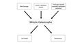 Mitotic Catastrophe Diagram 2.png 2 958 × 1 664; 192 KB
-
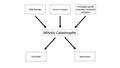 Mitotic Catastrophe Diagram version 2.png 2 958 × 1 664; 186 KB
-
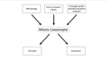 Mitotic Catastrophe Diagram.png 1 704 × 962; 99 KB
-
 Mitotic checkpoint vertebrates.png 680 × 501; 18 KB
-
 Mitotic LLC-PK1 cells, fluorescence microscopy (23700644352).jpg 1 800 × 1 200; 1,6 MB
-
 Network picture.png 346 × 259; 56 KB
-
 Neuroblast cell division - 486169.fig.002a.jpg 600 × 207; 10 KB
-
 Nondisjunction in Mitosis.jpg 479 × 473; 89 KB
-
 Normal and multipolar mitosis.tif 1 024 × 1 024; 3,14 MB
-
 Nuclear envelope breakdown and reassembly in mitosis.jpg 1 200 × 790; 142 KB
-
 Onion cells under a microscope.jpg 2 268 × 4 032; 1,52 MB
-
 Onion root cells.png 965 × 482; 1 012 KB
-
 Pair-pull-part mitosis and meiosis.png 1 021 × 716; 295 KB
-
 Prophase diagram.svg 512 × 407; 46 KB
-
 PSM V71 D104 Cells from the mouth of the salamander to show mitosis.png 1 387 × 2 060; 421 KB
-
 Remak cell1.jpg 256 × 192; 11 KB
-
RPE Cell Taxol treatment - mitotic catastrophe.webm 8,9 s, 654 × 480; 1,8 MB
-

-
 Securin phosphorylation sites2.jpg 407 × 198; 62 KB
-
 Securin-separase conserved.png 345 × 258; 77 KB
-
 Simplified network controls mitotic exit.jpg 960 × 633; 55 KB
-
 Spindle Assembly Checkpoint.png 1 916 × 1 051; 306 KB
-
 Spindle checkpoint vertebrates - en.png 960 × 720; 75 KB
-
 Stages of ciliate conjugation.svg 1 146 × 863; 116 KB
-
 Stages of early mitosis in a vertebrate cell (hy).svg 512 × 239; 69 KB
-
 Stages of early mitosis in a vertebrate cell with micrographs of chromatids.svg 512 × 1 170; 458 KB
-
 Stages of early mitosis in a vertebrate cell.svg 512 × 1 408; 156 KB
-
 Stages of late M phase in a vertebrate cell.svg 512 × 1 593; 211 KB
-
 StevensNM-StSp-1905-Pl-6-R12.jpg 1 336 × 578; 87 KB
-
 StevensNM-StSp-1905-Pl-6-R34.jpg 1 336 × 586; 98 KB
-
 StevensNM-StSp-1905-Pl-6-R56.jpg 1 336 × 637; 96 KB
-
 StevensNM-StSp-1905-Pl-6-R70.jpg 1 336 × 321; 54 KB
-
 Telophase during Mitosis.svg 512 × 384; 55 KB
-
 The events of the eukaryotic cell cycle.svg 512 × 256; 1,28 MB
-
 The twin brothers.tif 10 836 × 10 011; 21,31 MB
-
 Three cell growth types.png 499 × 507; 170 KB
-
 Trije tipi celične delitve.svg 694 × 700; 235 KB
-
 Tripolar Mitosis - breast carcinoma.jpg 2 048 × 1 536; 1,41 MB
-
 Tripolar Mitosis - bronchial wash.jpg 1 238 × 921; 365 KB
-
 Twelve sketches illustrating all successive stages of mitosis (23129542051).jpg 1 925 × 1 772; 1,26 MB
-
 Types of mitosis.png 664 × 448; 121 KB
-
 Whole Genome Doubled Instability.png 4 431 × 1 319; 667 KB
-
 Wilson1900Fig1.jpg 1 388 × 1 201; 296 KB
-
 Zellsubstanz-Kern-Kerntheilung.jpg 306 × 434; 36 KB
-
 Événements majeurs de la Mitose es.png 460 × 167; 54 KB
-
 Митоза (анимација).gif 865 × 579; 3,01 MB


.png)
.png)
.png)
.png)


.jpg)


.jpg)
.jpg)


.png)


.jpg)
.jpg)


.png)


.jpg)


.svg)


.jpg)


.gif)
.jpg)
.png)