Category:Placenta
Vai alla navigazione
Vai alla ricerca
organo temporaneo presente nelle femmine di alcuni mammiferi durante la gravidanza  | |||||
| Carica un file multimediale | |||||
| Audio della pronuncia | |||||
|---|---|---|---|---|---|
| Istanza di |
| ||||
| Sottoclasse di |
| ||||
| Aspetto di | |||||
| |||||
Sottocategorie
Questa categoria contiene le 7 sottocategorie indicate di seguito, su un totale di 7.
Pagine nella categoria "Placenta"
Questa categoria contiene un'unica pagina, indicata di seguito.
P
File nella categoria "Placenta"
Questa categoria contiene 84 file, indicati di seguito, su un totale di 84.
-
 201405 placenta.png 400 × 400; 66 KB
-
 A system of obstetrics (1888) (14592419007).jpg 1 568 × 1 160; 284 KB
-
 A system of obstetrics (1888) (14756113456).jpg 1 448 × 3 460; 790 KB
-
 A system of obstetrics (1888) (14776393984).jpg 2 148 × 3 428; 764 KB
-
 A system of obstetrics (1888) (14776758174).jpg 2 152 × 3 080; 737 KB
-

-
 A textbook of obstetrics (1898) (14594077878).jpg 1 752 × 2 852; 762 KB
-
 A textbook of obstetrics (1898) (14780390392).jpg 1 296 × 1 272; 307 KB
-
 Adrianus Spigelius, De formato foetu Wellcome L0026175.jpg 1 132 × 1 654; 990 KB
-
 Amnion.jpg 1 415 × 1 303; 1,16 MB
-

-
 An American text-book of physiology (1897) (14760365116).jpg 936 × 1 566; 551 KB
-

-
 Bidloo Ontleding 1690 57.jpg 1 200 × 1 618; 290 KB
-
 Bilobate Placenta with Velamentous Insertion of the Cord (8271783612).jpg 1 936 × 2 592; 1,31 MB
-
 Bilobate Placenta with Velamentous Insertion of the Cord.jpg 2 592 × 1 936; 1,31 MB
-
 C2orf72 Orthologs List.png 807 × 616; 305 KB
-
 Classification of placenta.jpg 800 × 354; 70 KB
-
 Cord & Placenta.jpg 1 200 × 1 600; 431 KB
-
 Diamniotic-dichorionic twin placenta - low mag.jpg 2 400 × 3 600; 4,61 MB
-
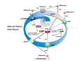
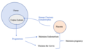
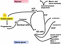 Early hormonal interaction after implantation.jpg 1 663 × 1 189; 125 KB
-
 Elements of the comparative anatomy of vertebrates (1886) (21057027940).jpg 1 328 × 1 500; 342 KB
-
-

-
 Fatty Acid Transport Mechanism.png 1 190 × 872; 534 KB
-

-

-
 Fetal circulation.jpg 3 600 × 3 591; 7,13 MB
-
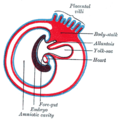
 FetalMembranes1L.jpg 424 × 400; 57 KB
-
 Feto-maternal interface showing uterine milk.jpg 2 994 × 1 475; 458 KB
-
 Fetus and placenta - journal.pbio.0060312.g001.jpg 1 072 × 1 072; 146 KB
-
 Film of placenta, donated by volunteer woman for research.jpg 2 560 × 1 440; 473 KB
-
 Final da implantação do blastocisto.png 599 × 506; 376 KB
-
 Foetus cat.jpg 667 × 827; 532 KB
-
 Gray0026-horz.jpg 960 × 480; 179 KB
-
 Gray26.svg 344 × 345; 21 KB
-
 Gray27.png 300 × 298; 10 KB
-
 Gray28.svg 396 × 455; 14 KB
-
 Gray39 cs.png 576 × 384; 50 KB
-
 Gray39 Placenta-hu.PNG 500 × 384; 69 KB
-
 Gray39 sp.PNG 500 × 384; 38 KB
-
 Gray39.png 500 × 384; 41 KB
-
 Haultain and Ferguson - placenta.svg 2 285 × 1 130; 3,36 MB
-
 HCG.Image.png 860 × 544; 62 KB
-
 Healthy Placenta.jpg 3 024 × 4 032; 2,98 MB
-

-

-
 Implantação do blastocisto.jpg 1 098 × 468; 263 KB
-
 Innesteling blastula.jpg 2 236 × 954; 700 KB
-
 Jenty; anatomy of the womb and foetus Wellcome L0030023.jpg 1 180 × 1 848; 1,13 MB
-
 Plate from "De formato foetu..." Fabricius, 1604 Wellcome L0008418.jpg 1 135 × 1 662; 983 KB
-
 Plate from "De formato foetu..." Fabricius, 1604 Wellcome L0008419.jpg 1 138 × 1 678; 995 KB
-
 LAENNNEC.jpg 4 011 × 3 007; 1,59 MB
-
 Lotus Birth Placenta.png 287 × 215; 108 KB
-
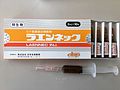
 MELSMON (Pharmaceuticals made from Placenta).jpg 3 895 × 2 309; 1,33 MB
-
Motion-and-Flexibility-in-Human-Cytochrome-P450-Aromatase-pone.0032565.s011.ogv 56 s, 480 × 360; 3,85 MB
-
 Navel cord.jpg 2 048 × 1 361; 304 KB
-
 Placenta (PSF).png 1 325 × 1 331; 155 KB
-
 Placenta 001.jpg 2 048 × 1 536; 547 KB
-
 Placenta 002.jpg 960 × 720; 104 KB
-
 Placenta belonging to twins. Wellcome L0009816.jpg 1 084 × 1 701; 957 KB
-
 Placenta cartoon.png 569 × 564; 108 KB
-
 Placenta crop.jpg 500 × 500; 107 KB
-
 Placenta held.jpg 2 048 × 1 361; 308 KB
-
 Placenta i bossa amniòtica.1178.jpg 952 × 665; 85 KB
-
 Placenta next to foetus and Placenta adhering to womb. Wellcome L0009815.jpg 1 008 × 1 736; 916 KB
-
 Placenta slide.jpg 614 × 498; 177 KB
-
 PlacentaEsq.png 720 × 553; 479 KB
-
 Placental morphologies encountered in placental mammals.jpg 800 × 432; 42 KB
-
 Placentation endothéliochoriale et éphithéliochorale.jpg 3 394 × 934; 424 KB
-
 Planeten und Placenten.jpg 1 564 × 1 147; 197 KB
-
 Plazenta.png 500 × 384; 40 KB
-
 Removing the placenta and umbilical cord after birth Wellcome L0038257.jpg 3 012 × 4 038; 3,25 MB
-
 Sejrsskjorte DMR-168020 2000.jpg 1 518 × 2 000; 784 KB
-
 Tab III; Placenta within amneotic sac Wellcome L0064774.jpg 3 854 × 6 370; 6,92 MB
-
 Tab VII; Details from placenta and other parts Wellcome L0064778.jpg 3 842 × 6 376; 5,58 MB
-
 Telocytes-Fig 5.tif 2 056 × 672; 2,14 MB
-
 Terracotta votive offering in the shape of placenta, Roman, Wellcome L0058478.jpg 4 256 × 2 832; 1,59 MB
-
 The diagnosis and treatment of diseases of women (1907) (14597881628).jpg 1 272 × 1 432; 483 KB
-
 The evolution of the placental interface.jpg 800 × 565; 68 KB
-
 The Fatty acid transport system of human placenta.jpg 720 × 540; 82 KB
-
 The placenta is detached and ready to deliver.jpg 3 600 × 2 282; 3,18 MB
-
 The science and art of midwifery (1891) (14579734977).jpg 2 004 × 1 788; 708 KB
-
 Umbilical Region, the Cord, and the Placenta at Term.jpg 778 × 635; 577 KB

_(14592419007).jpg)
_(14756113456).jpg)
_(14776393984).jpg)
_(14776758174).jpg)
_(14775676334).jpg)
_(14594077878).jpg)
_(14780390392).jpg)


_(14767442322).jpg)
_(14760365116).jpg)
_represents_as_well_as_means_%22Birth%22_or_%22Celestial_Being%22_or_%22Divinity%22_or_%22Eternity%22_or_%22Heaven%22_or_%22Immortality%22_or_%22Placenta%22_or_%22Sky%22.jpg)

.jpg)


_(21057027940).jpg)
.png)

_(14577869180).jpg)
_(14764250422).jpg)


.jpg)
_(14802956363).jpg)


.jpg)

.png)


_(14597881628).jpg)


_(14579734977).jpg)
