Category:Primitive streak
Jump to navigation
Jump to search
structure around day 15 of amniote embryogenesis | |||||
| Upload media | |||||
| Instance of |
| ||||
|---|---|---|---|---|---|
| Subclass of |
| ||||
| |||||
Media in category "Primitive streak"
The following 125 files are in this category, out of 125 total.
-
 2908 Germ Layers-02.jpg 1,781 × 1,590; 1.06 MB
-
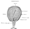
 A human embryo of 2 mm. in median sagittal section.jpg 838 × 1,006; 310 KB
-

-
-
-
 Agelena labyrinthica four stages in the development.jpg 988 × 691; 718 KB
-
 Agelena labyrinthica ventral plate at three stages.jpg 1,120 × 719; 454 KB
-
 Amphioxus Origin of the mesoderm.jpg 1,802 × 756; 793 KB
-

-
 Analysis of development (1955) (18166798962).jpg 1,012 × 1,504; 221 KB
-
-
-
-

-
 Aves Clustering of seemingly stochastic EMT underpins the formation of PS.jpg 780 × 1,500; 174 KB
-

-
 Aves Diagrams depicting the early stages of chick development.jpg 1,500 × 922; 152 KB
-
-
 Aves Dorsal view of a twenty-five-hour chick embryo with seven primitive segments.jpg 1,993 × 1,744; 2.21 MB
-
 Aves early pre-primitive streak blastoderm.jpg 822 × 523; 380 KB
-
-
 Aves EMT in the formation of the primitive streak.jpg 856 × 1,500; 351 KB
-
 Aves FGF signalling in mesoderm migration.jpg 2,152 × 612; 371 KB
-
 Aves gastrulation Mechanism of cell ingression.jpg 1,478 × 1,127; 175 KB
-
 Aves gastrulation Origins of flow organisation.jpg 1,660 × 1,579; 661 KB
-
 Aves gastrulation Tissue flows driving primitive streak formation.jpg 1,663 × 1,621; 710 KB
-
 Aves Median longitudinal section of a thirty-six-hour chick embryo.jpg 833 × 1,729; 271 KB
-

-

-
 Aves Schematic drawing of definitive endoderm origin. 12861 2007 233 MOESM3 ESM.tiff 1,500 × 1,500; 212 KB
-
 Aves Surface views of two stages of the blastoderm of the egg of the sparrow.jpg 1,456 × 789; 883 KB
-
 Aves Three stages of the blastoderm to show the extension of the mesoblast.jpg 1,486 × 626; 885 KB
-

-
-
 Aves View of the dorsal surface of a thirty-six-hour chick embryo.jpg 1,086 × 1,604; 918 KB
-
-
-
 C1. Primitive streak present (V03a).png 481 × 218; 46 KB
-
 Cell flow patterns during streak formation for different mechanisms.jpg 1,929 × 1,046; 1.7 MB
-

-
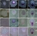
 Characterization of the SOX2T-positive territory of the epiblast in chicken embryo.jpg 2,113 × 1,581; 1.21 MB
-
-
 Didelphidae early development of blastoderm.jpg 1,045 × 788; 621 KB
-
 Dose-dependent reshaping of primitive streak.jpg 968 × 1,236; 743 KB
-
-

-
Dynamics of mesodermal cell ingression chicken embryo.ogv 12 s, 226 × 720; 4.2 MB
-
 EB1911 Peripatus - P. capensis - Series of Embryos.jpg 952 × 311; 78 KB
-
 Embryology (1949) (21285693065).jpg 1,035 × 1,115; 581 KB
-

-
-
 Formation of the primitive body plan following gastrulation in the mouse.png 1,279 × 1,187; 1,016 KB
-
 Formation of the primitive streak in the mammalian embryo.png 620 × 980; 584 KB
-
 Formation of the Primitive Streak.pdf 2,043 × 1,541; 14 KB
-
 Gastrulation forms in vertebrates.jpeg 1,280 × 1,173; 88 KB
-

-
 Gray13.png 215 × 245; 25 KB
-
 Gray13a.jpg 215 × 245; 31 KB
-
 Gray14.png 450 × 436; 25 KB
-
 Gray21.png 500 × 427; 24 KB
-
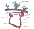
 Hand-book of physiology (1892) (14742414506).jpg 757 × 793; 565 KB
-
 Hydrophilus piceus embryos (01).jpg 754 × 750; 520 KB
-
 Kirkes' handbook of physiology (1907) (14769687492).jpg 1,091 × 918; 435 KB
-

-

-
-
-
-
-
-
-
-
-
-
-
-
-
-
Long-term tracking of a nuclear red stained chicken embryo.ogv 10 s, 248 × 718; 4.63 MB
-
Long-term tracking of the mesodermal progenitors chicken embryo.ogv 10 s, 236 × 432; 889 KB
-
Longevity of tracks along the primitive streak (PS) chicken embryo.ogv 5.9 s, 286 × 720; 1.48 MB
-
-

-

-
 Mesoderm-ring to -crescent transition.jpg 1,173 × 1,826; 1.01 MB
-
 Migration of epiblast cells in the mammalian embryo.png 866 × 530; 281 KB
-
Modeling-Gastrulation-in-the-Chick-Embryo-Formation-of-the-Primitive-Streak-pone.0010571.s002.ogv 17 s, 648 × 492; 16.95 MB
-
Modeling-Gastrulation-in-the-Chick-Embryo-Formation-of-the-Primitive-Streak-pone.0010571.s003.ogv 9.5 s, 700 × 350; 5.23 MB
-
Modeling-Gastrulation-in-the-Chick-Embryo-Formation-of-the-Primitive-Streak-pone.0010571.s004.ogv 21 s, 700 × 350; 17.02 MB
-
Modeling-Gastrulation-in-the-Chick-Embryo-Formation-of-the-Primitive-Streak-pone.0010571.s005.ogv 22 s, 1,050 × 350; 19.89 MB
-
Modeling-Gastrulation-in-the-Chick-Embryo-Formation-of-the-Primitive-Streak-pone.0010571.s006.ogv 24 s, 1,050 × 350; 14.22 MB
-
Modeling-Gastrulation-in-the-Chick-Embryo-Formation-of-the-Primitive-Streak-pone.0010571.s007.ogv 15 s, 700 × 700; 10.8 MB
-
Modeling-Gastrulation-in-the-Chick-Embryo-Formation-of-the-Primitive-Streak-pone.0010571.s008.ogv 30 s, 1,050 × 350; 22.29 MB
-
Modeling-Gastrulation-in-the-Chick-Embryo-Formation-of-the-Primitive-Streak-pone.0010571.s009.ogv 0.0 s, 1,056 × 352; 22.13 MB
-
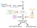
 Nodal signaling.jpg 1,168 × 1,198; 537 KB
-
 Number of TSOX2 double-positive cells during early chicken stages embryo.jpg 2,007 × 2,502; 828 KB
-

-
-
 Primitiv Node.jpg 720 × 504; 42 KB
-
-
 Quantification of the number of epiblast cells electroporated chicken embryo.jpg 730 × 1,687; 273 KB
-

-

-
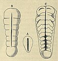 Scorpion ventral plate.jpg 624 × 653; 361 KB
-
-
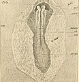
 Staging Human Gastrulation.jpg 3,493 × 3,010; 538 KB
-
 Sus domesticus embryos The primitive streak.jpg 1,123 × 912; 408 KB
-
 The Biological bulletin (20187079648).jpg 1,854 × 2,206; 1.25 MB
-
 The development of the chick; an introduction to embryology (1908) (20881489692).jpg 1,868 × 1,940; 1.26 MB
-
 The primitive streak and notochordal canal in Chelonia (Plate I) BHL4780730.jpg 2,577 × 1,849; 585 KB
-
 The primitive streak and notochordal canal in Chelonia (Plate II) BHL4780734.jpg 2,599 × 1,885; 3.31 MB
-
 The primitive streak and notochordal canal in Chelonia (Plate III) BHL4780738.jpg 2,104 × 1,877; 392 KB
-
 The primitive streak and notochordal canal in Chelonia (Plate IV) BHL4780742.jpg 2,054 × 3,526; 1.05 MB
-
 The primitive streak and notochordal canal in Chelonia (Plate V) BHL4780746.jpg 2,174 × 3,571; 849 KB
-
 The primitive streak and notochordal canal in Chelonia (Plate VI) BHL4780750.jpg 2,126 × 3,571; 1.01 MB
-
 The primitive streak and notochordal canal in Chelonia (Plate VII) BHL4780754.jpg 2,118 × 3,526; 778 KB
-
 The primitive streak and notochordal canal in Chelonia (Plate VIII) BHL4780758.jpg 2,555 × 1,863; 583 KB
-
 The primitive streak and notochordal canal in Chelonia (Plate X) BHL4780766.jpg 2,118 × 3,526; 1.49 MB
-
 The primitive streak and notochordal canal in Chelonia (Plate XI) BHL4780770.jpg 2,118 × 3,526; 1.13 MB
-
-
Tracking cell division time in the SOX2T region chicken embryo.ogv 16 s, 388 × 720; 7.73 MB
-
Tracking descendants of the anterior primitive streak (PS) epiblast at 25 somites chicken embryo.ogv 30 s, 444 × 720; 10.99 MB
-
Tracking the fate of descendants of the anterior epiblast at 25 somites chicken embryo.ogv 12 s, 1,280 × 578; 6.39 MB
-
 Vespertilio murinus Median longitudinal section through the blastoderm.jpg 1,797 × 447; 440 KB
-
 WNTPathway.png 325 × 710; 24 KB


_(14596981329).jpg)


_epiblast_chicken_embryo.jpg)
_(18166798962).jpg)


_of_primitve_streak.jpg)


.png)


_(20661197932).jpg)


_epithelial_phenotype_during_development_chicken_embryo.jpg)
_(14781787612).jpg)
_(21285693065).jpg)
_signature_genes_in_chicken_and_mouse_embryos.jpg)


_(14742414506).jpg)
.jpg)
_(14769687492).jpg)


_progenitor_territories_results_in_NMPs_remaining_as_the_major_PS_remnant_in_the_tail_bud.jpg)


_and_median_sagittal_sections_(g_and_h)_using_the_rabbit_as_a_model.jpg)


_analysis_of_the_posterior_tissue_precursors_during_posterior_axis_formation_chicken_embryo.jpg)


.jpg)
_(20881489692).jpg)
_BHL4780730.jpg)
_BHL4780734.jpg)
_BHL4780738.jpg)
_BHL4780742.jpg)
_BHL4780746.jpg)
_BHL4780750.jpg)
_BHL4780754.jpg)
_BHL4780758.jpg)
_BHL4780766.jpg)
_BHL4780770.jpg)

_node_of_a_thirty-six-hour_chick_embryo.jpg)