Category:Super resolution microscopy
Jump to navigation
Jump to search
This category is for images taken by microscopical techniques that circumvent the Abbe Limit
Subcategories
This category has the following 2 subcategories, out of 2 total.
S
- STED microscopy (9 F)
Media in category "Super resolution microscopy"
The following 51 files are in this category, out of 51 total.
-
 3D Dual Color Super Resolution Microscopy Cremer 2010.png 3,486 × 1,280; 3 MB
-
 3D-SIM-1 NPC Confocal vs 3D-SIM detail.jpg 512 × 517; 94 KB
-
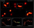
 3D-SIM-1 NPC Confocal vs 3D-SIM.jpg 1,069 × 848; 307 KB
-
 3D-SIM-2 Nucleus prophase 3d rotated.jpg 1,920 × 640; 377 KB
-
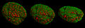
 3D-SIM-3 Prophase 3 color.jpg 902 × 896; 386 KB
-
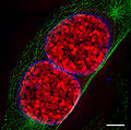
 3D-SIM-4 Anaphase 3 color.jpg 954 × 724; 264 KB
-
 ANNA-PALM.jpg 512 × 512; 71 KB
-
 Bacteria-3D-Double-Helix.jpg 4,956 × 5,310; 3.94 MB
-
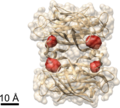 Biotin binding sites in streptavidin determined using COLD.png 872 × 785; 644 KB
-
Capture-of-microtubule-plus-ends-at-the-actin-cortex-promotes-axophilic-neuronal-migration-by-Video1.ogv 6.1 s, 885 × 765; 1.73 MB
-
-
Chromatin-Ring-Formation-at-Plant-Centromeres-Video1.ogv 6.3 s, 2,024 × 1,302; 2.46 MB
-
Chromatin-Ring-Formation-at-Plant-Centromeres-Video10.ogv 10 s, 2,024 × 1,302; 2.52 MB
-
Chromatin-Ring-Formation-at-Plant-Centromeres-Video11.ogv 17 s, 2,024 × 1,302; 7.51 MB
-
Chromatin-Ring-Formation-at-Plant-Centromeres-Video12.ogv 13 s, 2,090 × 1,272; 2.92 MB
-
Chromatin-Ring-Formation-at-Plant-Centromeres-Video3.ogv 17 s, 2,024 × 1,302; 8.74 MB
-
Chromatin-Ring-Formation-at-Plant-Centromeres-Video4.ogv 17 s, 2,024 × 1,302; 3.59 MB
-
Chromatin-Ring-Formation-at-Plant-Centromeres-Video8.ogv 29 s, 2,090 × 1,302; 6.3 MB
-
Chromatin-Ring-Formation-at-Plant-Centromeres-Video9.ogv 13 s, 2,154 × 1,302; 4.68 MB
-
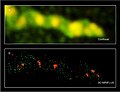
 FBALM DNA superresolution HeLa cell nucleus.png 3,568 × 1,508; 7.23 MB
-
 GFP Superresolution Christoph Cremer.JPG 538 × 389; 156 KB
-
-
-
-
-
-
-
-
-
-
 Label-free Localisation Microscopy SPDM - Super Resolution Microscopy Christoph Cremer.jpg 2,012 × 1,280; 1.55 MB
-
 Localization of a fluorophore using MINFLUX.png 3,672 × 2,118; 1.73 MB
-
 MINFLUX image of a hippocampal neuron.jpg 827 × 565; 71 KB
-
 MINFLUX image peroxisomes.jpg 828 × 672; 83 KB
-
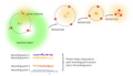
 MINFLUX localization.png 964 × 698; 111 KB
-
 MINFLUX principle (described in one dimension).png 2,026 × 2,130; 312 KB
-
 MINFLUX setup.jpg 595 × 842; 36 KB
-
 Opthalmology AMD Super Resolution Cremer.png 596 × 514; 271 KB
-
-
 Single YFP molecule superresolution microscopy-ru.png 340 × 686; 91 KB
-
 Single YFP molecule superresolution microscopy.png 340 × 686; 66 KB
-
 Storm super resolution.gif 1,000 × 343; 27.47 MB
-
Stormsuperresolution.webm 33 s, 1,000 × 343; 260 KB
-
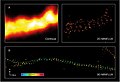
 Super-Resolution-Microtubules-BSC-1-cell.tif 1,439 × 566; 2.33 MB
-
-
-
-

-
 TUD nanostructure.png 2,141 × 2,141; 267 KB
-
 Two-color MINFLUX image of mitochondria.jpg 827 × 632; 98 KB
-
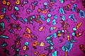 Микрокристаллы сульфата калия.JPG 5,472 × 3,648; 8.55 MB


.png)


