Category:White matter
Vai alla navigazione
Vai alla ricerca
part of the brain | |||||
| Carica un file multimediale | |||||
| Istanza di |
| ||||
|---|---|---|---|---|---|
| Sottoclasse di |
| ||||
| Parte di | |||||
| |||||
Sottocategorie
Questa categoria contiene le 8 sottocategorie indicate di seguito, su un totale di 8.
File nella categoria "White matter"
Questa categoria contiene 54 file, indicati di seguito, su un totale di 54.
-
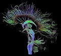
 3DPX-003145 Fractional Anisotrpy Tractography White Matter NevitDilmen.stl 5 120 × 2 880; 5,43 MB
-
 3DSlicer-KubickiJPR2007-fig6.jpg 714 × 296; 165 KB
-
 3DSlicer-Mislow-NeurosurgClinNAm2009-fig3.jpg 800 × 347; 198 KB
-
 3DSlicer-odonnell-miccai2006-fig2.jpg 1 386 × 652; 494 KB
-

-
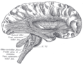
 A text-book of physiology for medical students and physicians (1911) (14755738306).jpg 1 352 × 1 160; 260 KB
-
-
 Arcuate fasciculus dissection and tractography.png 1 827 × 640; 1,55 MB
-
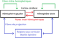 Classification des fibres de la substance blanche cérébrale.png 696 × 448; 18 KB
-
-
 Dissection of the human brain, Johann Christian Reil, 1812.jpg 1 451 × 1 598; 393 KB
-
 DTI Brain Tractographic Image Set.jpg 1 500 × 1 248; 921 KB
-
 DTI-sagittal-fibers.jpg 1 021 × 952; 294 KB
-
 Fiber tracts from six segments of the corpus callosum.gif 450 × 300; 46 KB
-

-
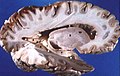
 Formalin-fixated human brain1.jpg 263 × 329; 128 KB
-
 Formalin-fixated human brain2.jpg 263 × 305; 119 KB
-
 Formalin-fixated human brain3.jpg 263 × 280; 94 KB
-
 Gray matter axonal connectivity.jpg 686 × 487; 147 KB
-
 Gray733.png 405 × 500; 58 KB
-
 Gray742 optical radiation.png 500 × 572; 193 KB
-
 Gray742.png 500 × 572; 57 KB
-
 Gray745.png 550 × 447; 62 KB
-
 Gray746.png 329 × 650; 21 KB
-
 Gray752.png 400 × 306; 30 KB
-
 Gray753.png 550 × 421; 54 KB
-
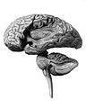 H. Mayo "Series of engravings", 1827; brain Wellcome L0015852.jpg 1 210 × 1 514; 373 KB
-

-

-
 Human brain right dissected lateral view description.JPG 653 × 413; 40 KB
-
 Human Brain.jpg 1 600 × 1 200; 658 KB
-
 Human cerebral cortex.png 411 × 384; 84 KB
-
 Lawrence 1960 2.21-23.png 2 024 × 2 840; 1,58 MB
-
 Lawrence 1960 2.25.png 1 932 × 1 380; 937 KB
-
 Meynert1885.PNG 1 400 × 867; 1,1 MB
-
 Mindmelding experiment.jpg 626 × 648; 100 KB
-
 Population-averaged human tractography atlas - Cranial Nerves.png 1 708 × 1 412; 1,17 MB
-

-
 Pre- and post-central gyrus, right hemisphere cropped.png 426 × 488; 140 KB
-
 Principles of Psychology (James) v1 p38.png 1 530 × 2 130; 878 KB
-
 PSM V35 D761 Direction of some of the fibers of the cerebrum.jpg 1 512 × 1 569; 407 KB
-
 PSM V46 D169 Course of the fibrous processes of the cortex.jpg 1 381 × 883; 303 KB
-
 Schematic illustration of projection fibers.jpg 1 300 × 1 114; 125 KB
-

-
 Simulated Connectivity Damage of Phineas Gage.png 1 302 × 1 817; 3,54 MB
-
 Sobo 1909 670-671.png 972 × 1 124; 3,13 MB
-
 Sobo 1909 672.png 984 × 1 167; 1,38 MB
-
 Sobo 1909 674.png 1 092 × 1 126; 3,52 MB
-
 Sobo 1909 675.png 2 292 × 2 052; 13,48 MB
-
 The Human Connectome.png 1 830 × 1 429; 5,78 MB
-
 Visible Human head slice.jpg 468 × 590; 70 KB
-
 White Matter Connections Obtained with MRI Tractography.png 336 × 463; 257 KB
-
 White matter fiber tracts.jpg 554 × 356; 51 KB
-
 White matter tracts.jpg 680 × 307; 123 KB


_(14767828014).jpg)
_(14755738306).jpg)


_in_cerebral_white_matter_(original).jpg)
_in_cerebral_white_matter.jpg)


_(14776972021).jpg)

_v1_p38.png)


_PMC3640224_figure_F3.jpg)
.jpg)