Category:Zoological Collection Rostock
Jump to navigation
Jump to search
Scientific university collection | |||||
| Upload media | |||||
| Instance of |
| ||||
|---|---|---|---|---|---|
| Location |
| ||||
| Street address |
| ||||
| official website | |||||
 | |||||
Subcategories
This category has only the following subcategory.
Media in category "Zoological Collection Rostock"
The following 77 files are in this category, out of 77 total.
-
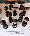
 Braun Max 1850 Rostock 1a.jpg 989 × 679; 388 KB
-
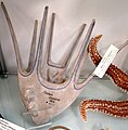
 Modell der Auricularia-Larve einer Holothurie (Seegurke) -Osterloh-.jpg 652 × 770; 155 KB
-
 Modell der Auricularia-Larve einer Holothurie (Seegurke) -Weisker-.jpg 525 × 1,068; 172 KB
-
 Modell der Bipinnaria-Larve von Asterias (Seestern) -Osterloh-.jpg 796 × 837; 199 KB
-
 Modell der Bipinnaria-Larve von Asterias (Seestern) -Weisker-.jpg 604 × 963; 154 KB
-
 Modell der Entwicklung des Frosches (Rana temporaria) -Ziegler 25- (2).jpg 1,000 × 1,217; 325 KB
-
 Modell der Entwicklung des Frosches (Rana temporaria) -Ziegler 25- (3).jpg 1,000 × 973; 260 KB
-
 Modell der Entwicklung des Frosches (Rana temporaria) -Ziegler 25- (4).jpg 1,000 × 643; 162 KB
-
 Modell der Entwicklung des Frosches (Rana temporaria) -Ziegler 25- (5).jpg 1,000 × 574; 175 KB
-
 Modell der Entwicklung des Frosches (Rana temporaria) -Ziegler 25- (6).jpg 1,000 × 670; 206 KB
-
 Modell der Entwicklung des Frosches (Rana temporaria) -Ziegler 25- (7).jpg 1,000 × 802; 173 KB
-
 Modell der Pluteus-Larve von Ophiuroidea (Schlangenstern) -Osterloh-.jpg 836 × 853; 241 KB
-
 Modell des Kopfes von Diphyllobothrium latum (Fischbandwurm) -Weisker-.jpg 498 × 1,805; 161 KB
-
 Modell des Kopfes von Taenia solium (Schweinebandwurm) -Weisker-.jpg 497 × 1,647; 147 KB
-
 Modell des Magens von Phoca vitulina (Seehund).jpg 1,000 × 990; 303 KB
-
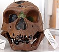 Modell des Schädels eines Neandertalers (La Chapelle-aux-Saints I) (3).jpg 1,000 × 879; 201 KB
-

-
 Modell eines dreiwurzeligen Backenzahns mit fortschreitender Karies (3).jpg 1,000 × 1,563; 229 KB
-
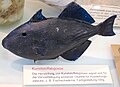 Modell eines Fischs.jpg 1,011 × 730; 226 KB
-
 Modell eines Ohrs - Gehörgangs (2).jpg 1,000 × 745; 207 KB
-
 Modell eines Ohrs - Gehörgangs.jpg 800 × 600; 80 KB
-
 Modell eines Schädels des Australopithecus africanus.jpg 1,000 × 1,137; 302 KB
-
 Modell eines Schädels des Australopithecus boisei.jpg 1,000 × 1,123; 255 KB
-

-
 Modell eines Schädels des Homo erectus modjokertensis (Java-Mensch).jpg 1,000 × 1,197; 216 KB
-
 Modell eines Schädels des Homo erectus rhodesiensis.jpg 1,000 × 1,220; 315 KB
-
 Modell eines Schädels des Homo steinheimensis (3).jpg 1,000 × 870; 298 KB
-
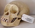 Modell eines Schädels des Pan troglodytes (Schimpanse).jpg 1,000 × 802; 205 KB
-

-
 Modell eines Schädels des Pan troglodytes (Schimpanse, weiblich).jpg 1,000 × 923; 224 KB
-
 Modell eines Schädels des Pongo pygmaeus (Borneo-Orang-Utan).jpg 1,000 × 1,002; 244 KB
-
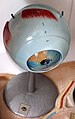 Modell vom menschlichen Auge.jpg 1,000 × 1,592; 292 KB
-
 Modell vom menschlichen Gehirn.jpg 625 × 768; 164 KB
-
 Modell vom menschlichen Herz (2).jpg 587 × 956; 184 KB
-
 Modell vom menschlichen Herz.jpg 539 × 768; 192 KB
-
 Modell vom menschlichen Torso -Louis M. Meusel- (2).jpg 1,000 × 1,383; 263 KB
-
 Modell vom menschlichen Torso -Louis M. Meusel-.jpg 1,000 × 1,945; 408 KB
-
 Modell vom Pantoffeltierchen.jpg 1,017 × 411; 126 KB
-
 Modell vom Trompetentierchen.jpg 467 × 1,416; 173 KB
-
 Modell von Bombyx mori (Seidenraupe) -Anatomische Lehrmittel Sonneberg-.jpg 1,000 × 700; 233 KB
-
 Modell von Cysticercus cellulasae (Schweinefinne) -Weisker-.jpg 794 × 547; 126 KB
-
 Modell von Ophiura (Schlangenstern).jpg 963 × 836; 224 KB
-
 Modell von Pulex irritans (weiblicher Menschenfloh) -Louis M. Meusel- (2).jpg 1,000 × 686; 138 KB
-
 Modell von Pulex irritans (weiblicher Menschenfloh) -Louis M. Meusel-.jpg 1,000 × 1,046; 276 KB
-
 Modell von Sinanthropus (Peking-Mensch) (2).jpg 800 × 600; 66 KB
-
 Modell von Sinanthropus (Peking-Mensch) (3).jpg 1,000 × 939; 231 KB
-
 Modell von Sinanthropus (Peking-Mensch).jpg 800 × 600; 66 KB
-
 Modell von Taenia multiceps (Quesenbandwurm) -Weisker-.jpg 499 × 694; 108 KB
-
 Modelle von Ascaris lumbricoides L. (Spulwurm) -Weisker-.jpg 1,000 × 733; 209 KB
-
-
 Modellreihe der Entwicklung des Froscheis (2).jpg 1,000 × 799; 184 KB
-
 Modellreihe der Entwicklung des Froscheis.jpg 1,000 × 1,084; 269 KB
-
 Modellreihe der Herzen von Wirbeltieren -SOMSO- (2).jpg 515 × 1,054; 150 KB
-
 Modellreihe der Herzen von Wirbeltieren -SOMSO- (3).jpg 2,736 × 3,648; 1.32 MB
-
 Modellreihe der Herzen von Wirbeltieren -SOMSO- (4).jpg 467 × 768; 97 KB
-
 Modellreihe der Herzen von Wirbeltieren -SOMSO- (5).jpg 521 × 1,025; 153 KB
-
 Modellreihe der Herzen von Wirbeltieren -SOMSO-.jpg 600 × 844; 135 KB
-
 Modellreihe der Kükenentwicklung im Ei bei Gallus domesticus (Haushuhn).jpg 1,734 × 400; 227 KB
-
 Modellreihe der Pluteus-Larve von Echinus (Seeigel) -Osterloh- (2).jpg 649 × 977; 161 KB
-
 Modellreihe der Pluteus-Larve von Echinus (Seeigel) -Osterloh- (3).jpg 638 × 1,039; 177 KB
-
 Modellreihe der Pluteus-Larve von Echinus (Seeigel) -Osterloh-.jpg 807 × 1,115; 218 KB
-
 Modellreihe der Pluteus-Larve von Ophiuroidea (Schlangenstern) -Weisker- (2).jpg 644 × 1,003; 157 KB
-

-
 Modellreihe der Pluteus-Larve von Ophiuroidea (Schlangenstern) -Weisker-.jpg 627 × 1,002; 150 KB
-
 Modellreihe von Wirbeltiergehirnen (2).jpg 1,000 × 472; 188 KB
-
 Modellreihe von Wirbeltiergehirnen (3).jpg 1,000 × 855; 212 KB
-
 Modellreihe von Wirbeltiergehirnen (4).jpg 1,000 × 981; 247 KB
-
 Modellreihe von Wirbeltiergehirnen.jpg 804 × 400; 145 KB
-
 Modellreihe zur Entwicklung des Hühnchens -Ziegler 26- (2).jpg 1,000 × 1,831; 369 KB
-
 Modellreihe zur Entwicklung des Hühnchens -Ziegler 26- (3).jpg 1,000 × 733; 230 KB
-
 Modellreihe zur Entwicklung des Hühnchens -Ziegler 26- (4).jpg 1,000 × 602; 184 KB
-
 Modellreihe zur Entwicklung des Hühnchens -Ziegler 26-.jpg 2,142 × 1,154; 1.51 MB
-
 Modellreihe zur Entwicklung des Lanzettfischchens -Ziegler 22- (28).jpg 1,000 × 628; 219 KB
-
 Modellreihe zur Entwicklung des Lanzettfischchens -Ziegler 22- (29).jpg 941 × 836; 152 KB
-
 Modellreihe zur Entwicklung des Lanzettfischchens -Ziegler 22- (30).jpg 1,000 × 687; 258 KB
-
 Modellreihe zur Entwicklung des Lanzettfischchens -Ziegler 22- (31).jpg 1,000 × 707; 242 KB
-
 Zoolog. Inst. HRO-20160411 195759.jpg 3,264 × 2,448; 2.29 MB

_-Osterloh-.jpg)
_-Weisker-.jpg)
_-Osterloh-.jpg)
_-Weisker-.jpg)
_-Ziegler_25-_(2).jpg)
_-Ziegler_25-_(3).jpg)
_-Ziegler_25-_(4).jpg)
_-Ziegler_25-_(5).jpg)
_-Ziegler_25-_(6).jpg)
_-Ziegler_25-_(7).jpg)
_-Osterloh-.jpg)
.jpg)
_(3).jpg)
.jpg)
.jpg)

.jpg)


.jpg)
.jpg)

.jpg)
.jpg)
_-Lehrmittelwerkst%C3%A4tten_des_Deutschen_Hygiene-Museums_Dresden-.jpg)
.jpg)
.jpg)


.jpg)

.jpg)

_-Anatomische_Lehrmittel_Sonneberg-.jpg)
_-Weisker-.jpg)
.jpg)
_-Louis_M._Meusel-_(2).jpg)
_-Louis_M._Meusel-.jpg)
_(2).jpg)
_(3).jpg)
.jpg)
_-Weisker-.jpg)
_-Weisker-.jpg)
_-Weisker-.jpg)
.jpg)

.jpg)
.jpg)
.jpg)
.jpg)

_-Osterloh-_(2).jpg)
_-Osterloh-_(3).jpg)
_-Osterloh-.jpg)
_-Weisker-_(2).jpg)
_-Weisker-_(3).jpg)
_-Weisker-.jpg)
.jpg)
.jpg)
.jpg)

.jpg)
.jpg)
.jpg)

.jpg)
.jpg)
.jpg)
.jpg)

_-Weisker-.jpg)
_-Weisker-.jpg)
.jpg)