Commons:Wiki Science Competition 2023/Microscopy images
Jump to navigation
Jump to search

Argentina · Estonia · Finland · France and Monaco · Indonesia · Ireland · Italy · Malaysia · Nigeria · North Macedonia · Poland · Russia · South Africa · Spain · Switzerland · Ukraine · United States ··· The rest of the World
People in Science · Microscopy images · Non-photographic media · Wildlife & nature · Astronomy images · General category · Image sets ··· Winners
Wiki Science Competition 2023 had 647 files submitted by 170 uploaders in the microscopy category.
Here are the finalists.
-
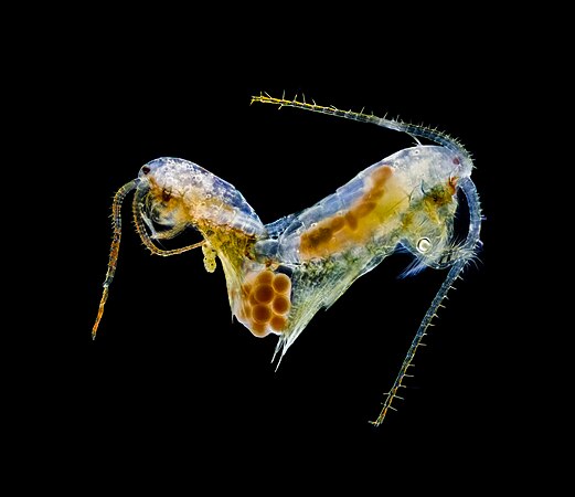 Copepods of the genus Limnocalanus mating. Dark field microscopy. Image taken during COVID19 pandemia. Photo by Antonio Segura
 -
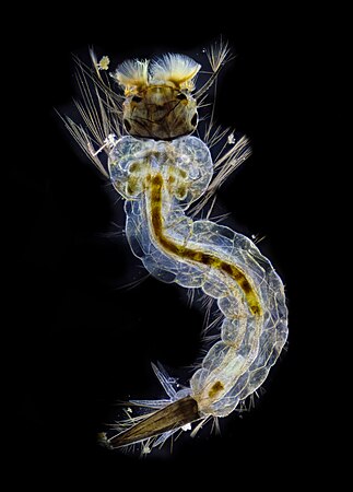 Mosquito larva seen with dark field and focus tracking. Image obtained during the COVID19 pandemia in Northern Patagonia, Argentina. Photo by Antonio Segura
 -
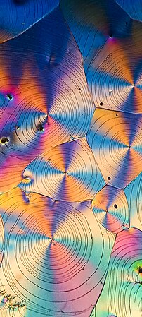 Ascorbic acid (Vitamin C) crystals seen under polarized light. Photo by Antonio Segura
 -
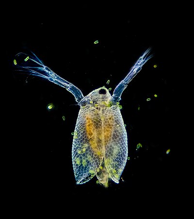
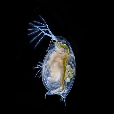 Daphnia with parthenogenetic eggs seen with dark field and focus tracking. 400x. Photo by Antonio Segura
 -
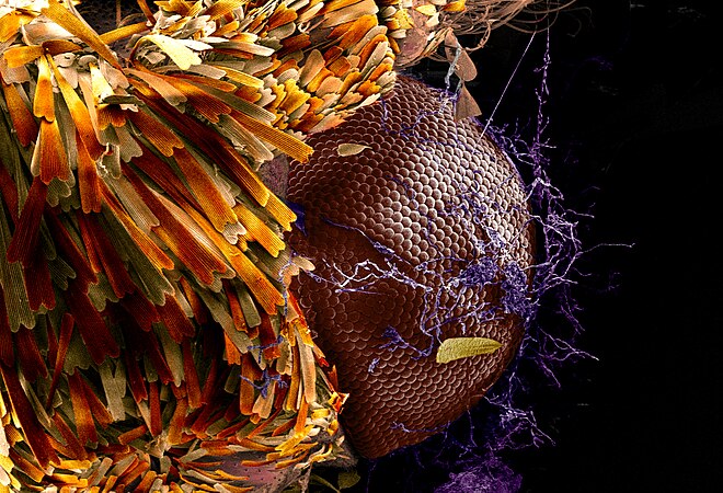 Mythimna loreyimima moth observed under a scanning electron microscope, digitally colored. A proliferation of Penicillium fungi is visible in the compound eye. Photo by Antonio Segura
 -
 Ceriodaphnia dubia seen with dark field, ventral view, focus tracking, 400x magnification. Photo by Antonio Segura
 -
 Entomopathogenic fungi Beauveria bassiana emerging from Tribolium castaneum. . Photo by Lautaro Preisegger, Daysi Espín Sánchez, and Carla Huarte-Bonnet
 -
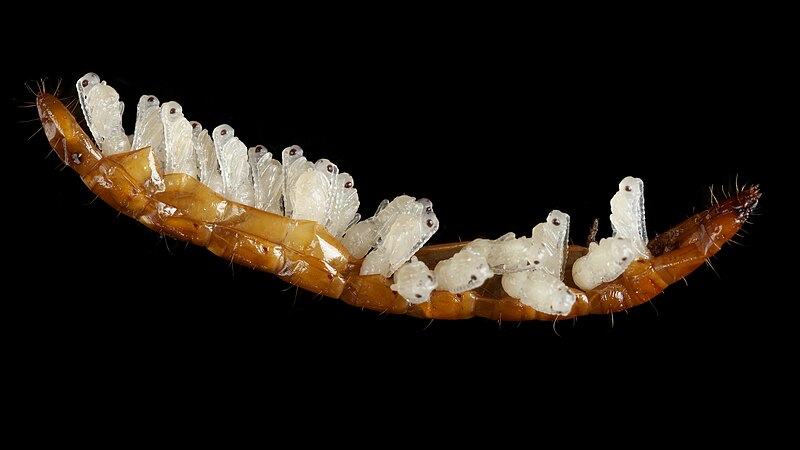 Pupae of the Paracodrus apterogynus in the shell remains of a click beetle (Elateridae) larva. The parasitized wireworm was collected in Estonia on August 27, 2021. Photo by Enno Merivee
 -
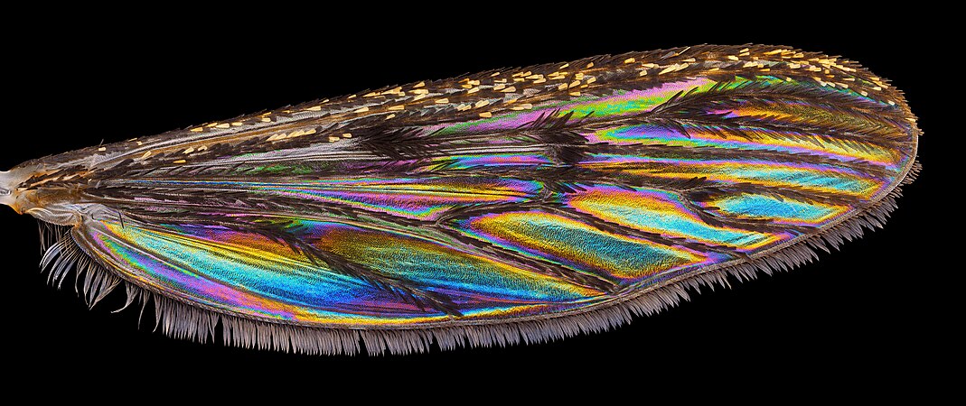 Recently, it was discovered that various insects with seemingly transparent wings, display vivid structural coloration against dark backgrounds. Wing interference patterns in the banded mosquito Culiseta annulata. Photo by Enno Merivee
 -
 Microscope image of colloid particles. Artificial opal made by drying a monodisperse sub-micrometer polystyrene sphere solution under a optical darkfield microscope. Photo by Siim Pikker
 -
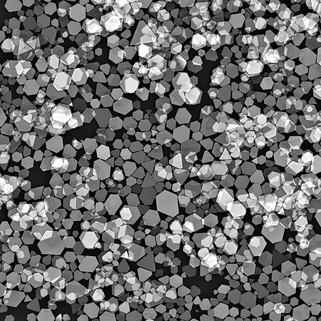 Silver micromirrors imaged with backscattered electrons in an electron microscope. Field of view 105 μm and imaging voltage 30 kV. These crystalline micromirrors are important model systems for micro and nanooptics research. Image by Siim Pikker
 -
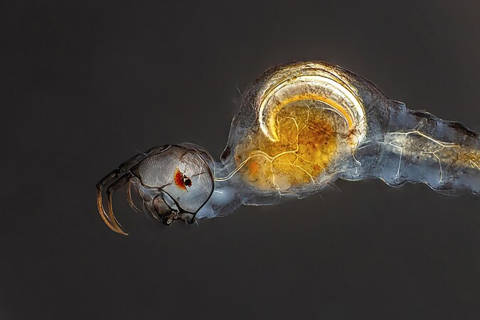 Chaoboridae larvae. Photo by Janek Lass
 -
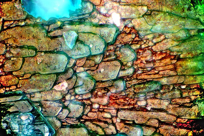 Oxidized surface of copper seen under a microscope. Sheet of copper was corroded, the bluish compound is copper(II) carbonate hydroxide. Photo by Tavo Romann
 -
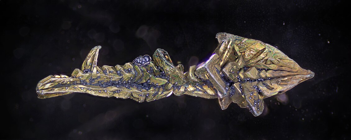 A vanadium crystal partially covered with purple (probably vanadium(IV) oxide) and green (some trivalent vanadium salts) corrosion products. Photo by Tavo Romann
 -
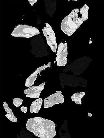 SEM image of zircon grains. These mineral grains were separated for radiometric dating, and they are about 100 µm thick. Image by Kallerna
 -
 Binocular magnifying glass photo of a corrosion pit (type I pit) on copper. This perforating corrosion concerns copper parts and is manifested by the presence of hemispherical craters surmounted by a pustule of greenish tint, under the pustule we find red Cu2O oxide. Photo by Kevin Ant 07
 -
 A blastula-stage sea urchin embryo. In turquoise we see the membranes of dividing cells, in red the microtubules of the mitotic spindle that act as "ropes" to pull the chromosomes, and in green the DNA in the form of chromosomes. Photo by AudeNommick
 -
 SEM image of ZnO nanorod structures showcasing the beauty of nanoworld taken from UKM research laboratory. Image by WMjay
 -
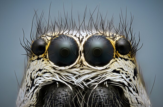 Eyes of Jumping spider (Salticus scenicus). Photo by Mikron86
 -
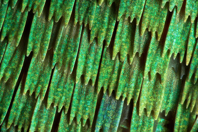 The structure of a butterfly's wing (Callophrys rubi), which consists of the so-called scales. Photo by Mikron86
 -
 A snowflake on a scale of 4:1. Photo by Mikron86
 -
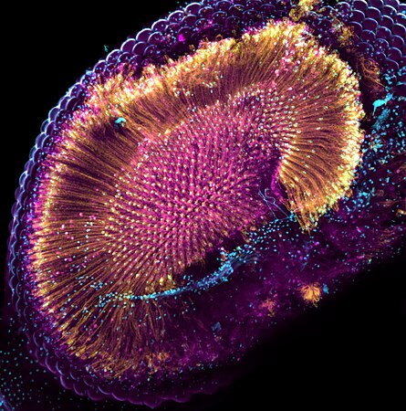 Confocal image of a fruit fly retina expressing a toxic form of the RdgB protein, leading to degeneration. Photo by Guillaume Thuery
 -
 A nucleation point in a thin crystalline film of an iron-based compound photographed through compensated polarisation microscopy. KarlGaff
 -
 Cells packed with chloroplasts and birefringence exhibted by the cell walls in a moss leaf. KarlGaff
 -
 This micrograph shows a vein of anhydrite (large triangular shapes) cutting a host rock of siltstone, filled with quartz and clay minerals. Photo by Aileen555
 -
 Onion cells under the microscope. Photo by Thepixiefrom02
 -
 Mullet leaf - Verbascum sp. observed under a microscope. Photo by Thepixiefrom02
 -
 Parasites in freshwater mussel Unio sp. under the binocular. Photo by Dijana Blazekovic
 -
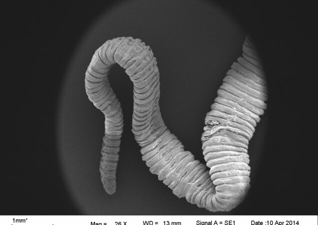 Bothriocephalus opsariichthydis. SEM image by Dijana Blazekovic
 -
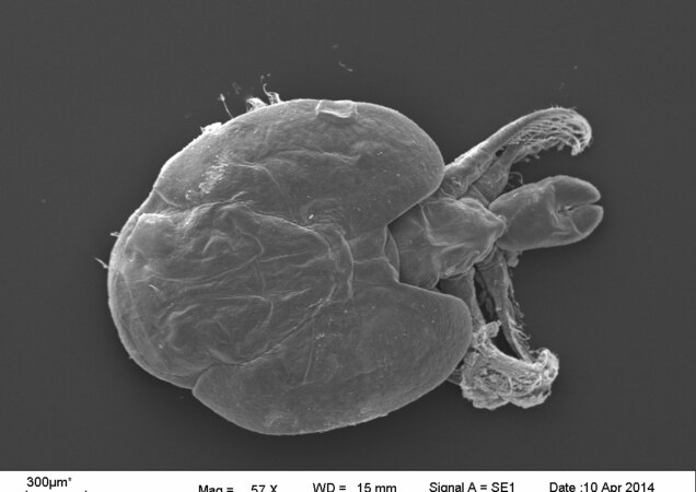
 Argulus foliaceus. SEM image by Dijana Blazekovic
 -
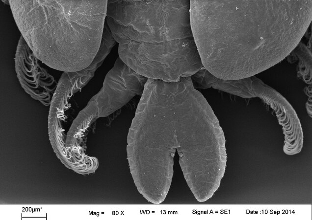
 Dorsal part of Argulus foliaceus. SEM image by Dijana Blazekovic
 -
 Microcrystals of Acetylsalicylic acid under an optical microscope at 60x between linear polarizing filters. Photo by Piero Stroppa
 -
 Photomicrography of Cymbella peraspera diatom, from a sample taken on 2022-11-15 in Valle dei Mulini, Vicenza, Italy. Photo by ExaVolt
 -
 Detail of the oral membranelles of Euplotes vanleeuwenhoeki under a scanning electron microscope. Photo by V.Serra
 -
 A cell culture (COS-7) colored with phalloidin-fitc (phalloidin conjugated to fluorescein diluted in PBS). Photo by Bioguy10
 -
 Image was taken with an optical microscope at 40x of two conidiophores of Aspergillus niger, formed by a globose vesicle. Photo by Chiara Marraccini
 -
 Microcrystals of Ascorbic acid (Vitamin C) under an optical microscope at 60x between linear polarizing filters. Photo by Piero Stroppa
 -
 Two Strontium Titanate (SrTiO3) dendritic crystals. Photo by Maria Teresa Buscaglia


_vistos_con_lu_polarizada.jpg)


