File:Fluorescence Imaging 02.jpg
Jump to navigation
Jump to search
Size of this preview: 800 × 534 pixels. Other resolutions: 320 × 213 pixels | 640 × 427 pixels | 928 × 619 pixels.
Original file (928 × 619 pixels, file size: 187 KB, MIME type: image/jpeg)
Captions
Captions
Add a one-line explanation of what this file represents
| Description |
Deutsch: Fluoreszenzbildgebung eines orthotopen Implantates eines Pankreaskarzinoms eine Maus. Die Bildgebung dient zu Bestimmung des Tumorvolumens. Die Maus wurde zwei Wochen nach der Injektion von humanen Pankreastumorzellen vom Typ XPA-1 fotografiert. Als Farbstoff wurde rot-fluoreszierendes Protein verwendet (RFP). Die Bildreihe A-C zeigt die betäubte Maus. Bild A ist ein Fusionsbild aus sichtbarem Licht und Fluoreszenzaufnahme. Bild B zeigt die Fluoreszenz des mit RFP markierten Tumors (nicht quantitativ). Bild C ist eine monochrome quantitative Aufnahme der Fluoreszenz des Tumors aus Bild A+B. In Reihe D-F wurde die Bauchdecke der Maus geöffnet und die Aufnahmen von A biC wiederholt. Deutlich zu erkennen ist die bessere Auflösung der Bilder, da die störenden Einflüsse der Bauchdecke fehlen.
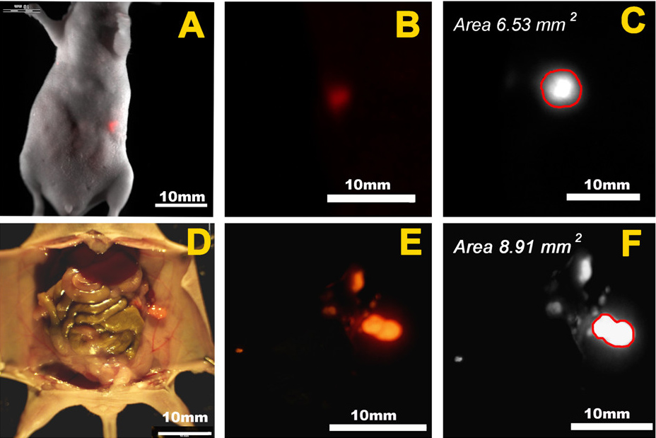
English: Fluorescence imaging of orthotopic pancreatic tumor implants and measurement of tumor size. This mouse was imaged two weeks after orthotopic injection of a human pancreatic cell line (XPA-1) stably expressing red fluorescent protein (RFP). A-C. Evaluation of tumor size by whole body imaging of an anesthetized mouse. A. Bright field and color fluorescence images were overlaid to show the coronal image of the fluorescent tumor. B. Enlarged whole body fluorescence image of tumor. This image was obtained using a color (non-quantitative) fluorescence imaging camera. C. Monochrome (quantitative) fluorescence image of the tumor shown in panels A and B. This coronal image was analyzed using ImageJ software to determine the cross-sectional size of the tumor in millimeters squared (mm2). D-F. Evaluation of tumor size at necropsy. D. Overlay of bright field and color fluorescence images showing the fluorescent tumor implant at necropsy. E. Enlarged color fluorescence-only image of the tumor as shown in D. F. As for panel C above, the cross-sectional size of the tumor shown in panels D and E was determined using ImageJ software. |
| Date | published 8 April 2009 |
| Source | Cynthia S Snyder, Sharmeela Kaushal, Yuko Kono, Hop S Tran Cao, Robert M Hoffman and Michael Bouvet: Complementarity of ultrasound and fluorescence imaging in an orthotopic mouse model of pancreatic cancer. In: BMC Cancer 2009, 9:106 doi:10.1186/1471-2407-9-106 Open Access |
| Author | Cynthia S Snyder, Sharmeela Kaushal, Yuko Kono, Hop S Tran Cao, Robert M Hoffman and Michael Bouvet |
I, the copyright holder of this work, hereby publish it under the following license:
This file is licensed under the Creative Commons Attribution-Share Alike 2.0 Generic license.
- You are free:
- to share – to copy, distribute and transmit the work
- to remix – to adapt the work
- Under the following conditions:
- attribution – You must give appropriate credit, provide a link to the license, and indicate if changes were made. You may do so in any reasonable manner, but not in any way that suggests the licensor endorses you or your use.
- share alike – If you remix, transform, or build upon the material, you must distribute your contributions under the same or compatible license as the original.
File history
Click on a date/time to view the file as it appeared at that time.
| Date/Time | Thumbnail | Dimensions | User | Comment | |
|---|---|---|---|---|---|
| current | 20:07, 22 June 2010 | | 928 × 619 (187 KB) | Kuebi (talk | contribs) | {{Information |Description={{de|Fluoreszenzbildgebung eines orthotopen Implantates eines Pankreaskarzinoms eine Maus. Die Bildgebung dient zu Bestimmung des Tumorvolumens. Die Maus wurde zwei Wochen nach der Injektion von humanen Pankreastumorzellen vom T |
You cannot overwrite this file.
File usage on Commons
There are no pages that use this file.
File usage on other wikis
The following other wikis use this file:
- Usage on de.wikipedia.org