File:SI organoid 1.tif
Jump to navigation
Jump to search

Size of this PNG preview of this TIF file: 800 × 592 pixels. Other resolutions: 320 × 237 pixels | 640 × 474 pixels | 1,024 × 758 pixels | 1,280 × 948 pixels | 2,604 × 1,928 pixels.
Original file (2,604 × 1,928 pixels, file size: 19.16 MB, MIME type: image/tiff)
Captions
Captions
Budding Mini Gut
Summary[edit]
| Description |
English: Immunofluorescence image of intestinal organoids generated using small intestine from ApcMin/+ mice, which carry an Apc mutation leading to the spontaneous development of intestinal tumours. Cells are stained for Protein kinase B, also known as AKT (red), epithelial cadherin (green) and nucleus (blue). This image was obtained using a Leica microscope with THUNDER Imaging Systems (20x objective). Multiple tile images and Z stacks were captured to cover the whole organoid. |
| Date | |
| Source | Own work |
| Author | Abhimanu.pandey |
Licensing[edit]
I, the copyright holder of this work, hereby publish it under the following license:
This file is licensed under the Creative Commons Attribution 4.0 International license.
- You are free:
- to share – to copy, distribute and transmit the work
- to remix – to adapt the work
- Under the following conditions:
- attribution – You must give appropriate credit, provide a link to the license, and indicate if changes were made. You may do so in any reasonable manner, but not in any way that suggests the licensor endorses you or your use.
| This image was uploaded as part of Wiki Science Competition 2019. |
This image has been assessed using the Quality image guidelines and is considered a Quality image.
|
File history
Click on a date/time to view the file as it appeared at that time.
| Date/Time | Thumbnail | Dimensions | User | Comment | |
|---|---|---|---|---|---|
| current | 00:19, 7 December 2019 | 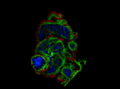 | 2,604 × 1,928 (19.16 MB) | Abhimanu.pandey (talk | contribs) | User created page with UploadWizard |
You cannot overwrite this file.
File usage on Commons
The following 7 pages use this file:
- User talk:Abhimanu.pandey
- Commons:Quality images/Subject/Microscopic
- Commons:Quality images candidates/Archives June 13 2021
- Commons:Wiki Science Competition 2019/Winners/Australia
- Commons:Wiki Science Competition 2019/Winners/Microscopy images
- Commons:Wiki Science Competition 2019/Winners/Microscopy images/round 2
- Commons:Wiki Science Competition 2019 in Australia
File usage on other wikis
The following other wikis use this file:
- Usage on nl.wikipedia.org