File:The Dental cosmos (1890) (14778827752).jpg
From Wikimedia Commons, the free media repository
Jump to navigation Jump to search

Original file(1,852 × 2,858 pixels, file size: 893 KB, MIME type: image/jpeg)

Captions

Captions

Add a one-line explanation of what this file represents

Summary[edit]

Description
English:

Identifier: dentalcosmos3218whit (find matches)
Title: The Dental cosmos
Year: 1890 (1890s)
Authors: White, J. D McQuillen, J. H. (John Hugh), 1826-1879 Ziegler, George Jacob, 1821-1895 White, James William, 1826-1891 Kirk, Edward C. (Edward Cameron), 1856-1933 Anthony, L. Pierce (Lovick Pierce), b. 1877
Subjects: Dentistry Dentistry
Publisher: Philadelphia, S. S. White Dental Manufacturing Co
Contributing Library: Yale University, Cushing/Whitney Medical Library
Digitizing Sponsor: The College of Physicians of Philadelphia and the National Endowment for the Humanities

View Book Page: Book Viewer
About This Book: Catalog Entry
View All Images: All Images From Book
Click here to view book online to see this illustration in context in a browseable online version of this book.

Text Appearing Before Image:
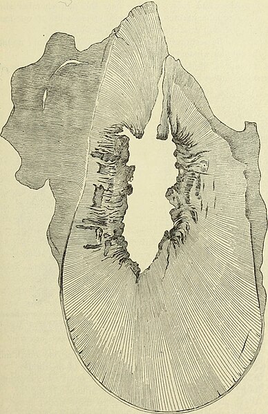
infiltration what-ever. (See Fig. 1.) No. 5. Section near open end decayed ; section near apex showsabout one-fourth of the canals infected with straggling cocci to a depthof one-fourth to one-half millimeter. No. 6. The end section is slightly decayed and correspondingly in-fected. The section near the apex is protected on one side by tube-less dentine ; on the other side eighteen tubules are infected by a fewstraggling bacilli to a depth of one-half to three-fourths millimeter(Fig. 3). A few of the tubules contain also cocci (Fig. 3, a, d), fromwhich we must conclude either that the dentine is infected with twokinds of bacteria or with a pleomorphous bacterium. No. 7. The root in this case contained an inflamed pulp ; no traceof tubular infection is anywhere to be discovered. DECOMPOSITION OF CONTENTS OF DENTINAL TUBULES, ETC. 353 No. 8. Section from about middle of root ; twenty-six of the tubulesare infected with cocci to a depth of about one-tenth millimeter. (SeeFig. 4-) Fig. 2.
Text Appearing After Image:
Fig. 2.—Cross-section of the posterior root of a lower molar, showing the advance of decayfrom the canal. 20 : 1. No. 9. The sections show again that the canal is in part lined withmodified dentine. In two or three tubules only was I able to findabout a dozen bacteria. The root was abscessed.vol. xxxii.—26 354 THE DENTAL COSMOS. No. 10. The canal was found filled with masses of bacteria (seeFig. 5), but no infection of the tubules worth mentioning could bedetected, only a few cocci being visible in three or four tubules. Thecanal was again partly lined with modified dentine. Root abscessed. No. 11. Abscessed root. Section near the pulp-chamber showsnearly all the tubules infected to the depth of one-tenth to one-fourthmillimeter. Section near apex free from infection. No. 12. Ground section from near the middle of the root ; no infec-tion of the tubules whatever. Nos. 13 and 14. Both partially protected by modified dentine, andboth free from infection. Fig. 3. Fig. 4. IS ijriili a

Note About Images

Please note that these images are extracted from scanned page images that may have been digitally enhanced for readability - coloration and appearance of these illustrations may not perfectly resemble the original work.
Date
Source

https://www.flickr.com/photos/internetarchivebookimages/14778827752/

Author

White, J. D; McQuillen, J. H. (John Hugh), 1826-1879; Ziegler, George Jacob, 1821-1895; White, James William, 1826-1891; Kirk, Edward C. (Edward Cameron), 1856-1933;

Anthony, L. Pierce (Lovick Pierce), b. 1877
Permission
(Reusing this file)
At the time of upload, the image license was automatically confirmed using the Flickr API. For more information see Flickr API detail.
Volume
InfoField
1890
Flickr tags
InfoField
Flickr posted date
InfoField
29 July 2014


Licensing[edit]

This image was taken from Flickr's The Commons. The uploading organization may have various reasons for determining that no known copyright restrictions exist, such as:
  1. The copyright is in the public domain because it has expired;
  2. The copyright was injected into the public domain for other reasons, such as failure to adhere to required formalities or conditions;
  3. The institution owns the copyright but is not interested in exercising control; or
  4. The institution has legal rights sufficient to authorize others to use the work without restrictions.

More information can be found at https://flickr.com/commons/usage/.


Please add additional copyright tags to this image if more specific information about copyright status can be determined. See Commons:Licensing for more information.
This image was originally posted to Flickr by Internet Archive Book Images at https://flickr.com/photos/126377022@N07/14778827752. It was reviewed on 15 September 2015 by FlickreviewR and was confirmed to be licensed under the terms of the No known copyright restrictions.

15 September 2015

File history

Click on a date/time to view the file as it appeared at that time.

Date/TimeThumbnailDimensionsUserComment
current08:11, 15 September 2015Thumbnail for version as of 08:11, 15 September 20151,852 × 2,858 (893 KB) (talk | contribs)== {{int:filedesc}} == {{information |description={{en|1=<br> '''Identifier''': dentalcosmos3218whit ([https://commons.wikimedia.org/w/index.php?title=Special%3ASearch&profile=default&fulltext=Search&search=insource%3A%2Fdentalcosmos3218whit%2F find ma...

There are no pages that use this file.