File:Vancomycin resistance.svg

Original file (SVG file, nominally 2,103 × 720 pixels, file size: 1.4 MB)
Captions
Captions
Summary[edit]
| Description |
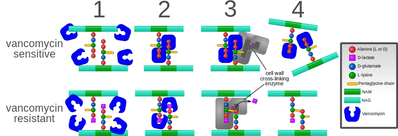
English: Diagram depicting the action of the antibiotic vancomycin and one way bacteria have evolved resistance to it.
Background: The bacterial cell wall consists of strands of repeating N-acetylglucosamine (NAG) and N-acetylmuramic acid (NAM) subunits. The NAM subunits have short peptide chains attached to them. (The exact composition of these can vary. The proximal alanine is usually L-ala and the distal two are usually D-ala.) These peptide chains are involved in forming cross-links between the strands of the cell wall. These cross-links are essential to a functioning cell wall. 1. Vancomycin is added to the bacterial environment while it is trying to synthesize new cell wall. Here, the cell wall strands have been synthesized, but not yet cross-linked. 2. Vancomycin recognizes and binds to the two D-ala residues on the end of the peptide chains. However, in resistant bacteria, the last D-ala residue has been replaced by a D-lactate, so vancomycin cannot bind. 3. In resistant bacteria, cross-links are successfully formed. However, in the non-resistant bacteria, the vancomycin bound to the peptide chains prevents them from interacting properly with the cell wall cross-linking enzyme. 4. In the resistant bacteria, stable cross links are formed. In the sensitive bacteria, cross-links cannot be formed and the cell wall falls apart. |
| Date | |
| Source | Own work |
| Author | Mcstrother |
| Other versions |
[]
File:Vancomycin resistance.svg has 0 translations. |


Licensing[edit]
- You are free:
- to share – to copy, distribute and transmit the work
- to remix – to adapt the work
- Under the following conditions:
- attribution – You must give appropriate credit, provide a link to the license, and indicate if changes were made. You may do so in any reasonable manner, but not in any way that suggests the licensor endorses you or your use.
File history
Click on a date/time to view the file as it appeared at that time.
| Date/Time | Thumbnail | Dimensions | User | Comment | |
|---|---|---|---|---|---|
| current | 18:27, 3 October 2023 | 2,103 × 720 (1.4 MB) | Santanyiner (talk | contribs) | File uploaded using svgtranslate tool (https://svgtranslate.toolforge.org/). Added translation for ca. | |
| 02:08, 10 September 2011 | 2,103 × 720 (1.65 MB) | Mcstrother (talk | contribs) | Updated to reflect correct mechanism of cross-linking. | ||
| 14:57, 3 May 2011 | 2,103 × 720 (1.65 MB) | Mcstrother (talk | contribs) | Changed fonts to Liberation Sans | ||
| 03:32, 10 April 2011 | 2,103 × 720 (1.65 MB) | Mcstrother (talk | contribs) | {{Information |Description ={{en|1=Diagram depicting the action of the antibiotic vancomycin and one way bacteria have evolved resistance to it. Background: The bacterial cell wall consists of strands of repeating N-acetylglucosamine (NAG) and N-acety |
You cannot overwrite this file.
File usage on Commons
The following 5 pages use this file:
File usage on other wikis
The following other wikis use this file:
- Usage on en.wikipedia.org
- Usage on he.wikipedia.org
- Usage on sv.wikipedia.org
- Usage on zh.wikipedia.org