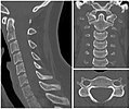
Scrollable computed tomography images of a normal brain (case 2)
Computer tomography of human brain, from base of the skull to top. Taken with intravenous contrast medium.
It was taken on 23 March 2007 on the uploader, after a 20-minute episode of homonymous hemianopsia with loss of the left visual field, but nothing strange was found. Three episodes of scotoma occurred in the following years, whereof the last one was scintillating (depiction). Otherwise, there were no further neurological symptoms.
To move through the images, hover over the image and roll a scroll wheel, drag the image up or down, or click the < or the > above each stack. This functionality should activate when the page is fully loaded, which may take some time. While scrolling, there may be flickering the first time each image appears in the stack.

.png)
.png)
.png)
.png)
.png)
.png)
.png)
.png)
.png)
.png)
.png)
.png)
.png)
.png)
.png)
.png)
.png)
.png)
.png)
.png)
.png)
.png)
.png)
.png)
.png)
.png)
.png)
.png)
.png)
.png)
.png)
.png)
.png)
.png)
Licensing[edit]
| This file is made available under the Creative Commons CC0 1.0 Universal Public Domain Dedication. | |
| The person who associated a work with this deed has dedicated the work to the public domain by waiving all of their rights to the work worldwide under copyright law, including all related and neighboring rights, to the extent allowed by law. You can copy, modify, distribute and perform the work, even for commercial purposes, all without asking permission.
|
All body locations[edit]
Available galleries of Public Domain scrollable computed tomography images of normal anatomy:
-
 Brain, case 1: No intravenous contrast.
 -
 Brain, case 2: contrast CT, axial plane only
 -
-
-
