Category:Media from National Cancer Institute Visuals Online with known IDs
Jump to navigation
Jump to search
This image was released by the National Cancer Institute, an agency part of the National Institutes of Health, with the ID [1] (image) (next).
This tag does not indicate the copyright status of the attached work. A normal copyright tag is still required. See Commons:Licensing.
|
Media in category "Media from National Cancer Institute Visuals Online with known IDs"
The following 77 files are in this category, out of 2,342 total.
(previous page) (next page)-
 Hands.jpg 3,200 × 2,133; 817 KB
-
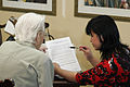 Woman reading.jpg 2,592 × 1,728; 2.43 MB
-
 Adult hands.jpg 2,592 × 1,728; 2.49 MB
-
 Woman's hands.jpg 2,592 × 1,728; 2.08 MB
-
 Woman (headshot).jpg 3,000 × 2,000; 898 KB
-
 Woman (headshot) (1).jpg 3,000 × 2,000; 848 KB
-
 Couple clasping hands.jpg 3,000 × 2,000; 714 KB
-
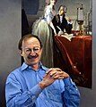 National Cancer Institute director Harold E. Varmus (6).jpg 938 × 1,054; 182 KB
-
 National Cancer Institute director Harold E. Varmus.jpg 1,008 × 1,589; 191 KB
-
 National Cancer Institute director Harold E. Varmus (1).jpg 3,008 × 2,000; 1.29 MB
-
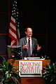 Town hall meeting - 2010.jpg 2,000 × 3,000; 853 KB
-
 Town hall meeting - 2010 (1).jpg 2,784 × 1,848; 517 KB
-
 Town hall meeting - 2010 (2).jpg 1,848 × 2,784; 738 KB
-
 National Cancer Institute director Harold E. Varmus (2).jpg 2,000 × 3,000; 521 KB
-
 National Cancer Institute director Harold E. Varmus (3).jpg 3,200 × 2,238; 541 KB
-
 National Cancer Institute director Harold E. Varmus (4).jpg 3,200 × 2,133; 574 KB
-
 National Cancer Institute director Harold E. Varmus (5).jpg 2,354 × 3,200; 1.15 MB
-
 Town hall meeting - 2010 (3).jpg 762 × 1,140; 136 KB
-
 Canellos, george.jpg 2,311 × 2,862; 619 KB
-
 Carter, stephen.jpg 2,252 × 2,843; 575 KB
-
 Cullen, william.jpg 2,067 × 2,836; 569 KB
-
 Flamm, william.jpg 2,833 × 2,280; 600 KB
-
 Friedell, gilbert.jpg 1,378 × 1,965; 159 KB
-
 Callan, laurence.jpg 2,254 × 2,831; 562 KB
-
 Former National Cancer Institute director Carl Baker (1970 - 1972) (1).jpg 2,280 × 2,833; 575 KB
-
 Baker, Carl and Rauscher, Frank.jpg 2,875 × 2,223; 546 KB
-
 Brace, kirkland.jpg 2,232 × 2,794; 415 KB
-
 Bruno, anthony.jpg 2,238 × 2,809; 573 KB
-
 Gordon, mordecai.jpg 2,833 × 2,232; 547 KB
-
 Haenszel, william.jpg 2,264 × 2,823; 366 KB
-
 Hammond, william.jpg 2,277 × 2,836; 562 KB
-
 Hartwell, jonathan.jpg 2,270 × 2,845; 460 KB
-
 Former National Cancer Institute director John Heller (1948 - 1960) (1).jpg 2,292 × 2,869; 295 KB
-
 Heston, walter.jpg 2,274 × 2,843; 349 KB
-
 Hirata, tadashi.jpg 2,286 × 2,847; 521 KB
-
 Holdenried, Robert.jpg 2,280 × 2,843; 662 KB
-
 Johnson, ralph.jpg 2,220 × 2,867; 556 KB
-
 Mardiney, michael.jpg 2,241 × 2,800; 386 KB
-
 Newell, guy.jpg 2,290 × 2,875; 346 KB
-
 Former National Cancer Institute director Frank Rauscher (1972 - 1976) (1).jpg 2,250 × 2,764; 403 KB
-
 Reed, C. D..jpg 2,229 × 2,884; 478 KB
-
 Former National Cancer Institute director Leonard Scheele (1947 - 1948) (1).jpg 2,280 × 2,845; 395 KB
-
 NCI campus at shady grove.jpg 3,000 × 2,105; 1.99 MB
-
 NCI campus at shady grove (1).jpg 3,000 × 2,028; 1.78 MB
-
 NCI campus at shady grove (2).jpg 3,000 × 1,882; 1.79 MB
-
 Lasers used for computed tomography positioning.jpg 3,000 × 2,000; 5.63 MB
-
 Preparing genotyping arrays.jpg 3,000 × 2,000; 2.48 MB
-
 Women look over publication (1).jpg 3,000 × 2,000; 6.2 MB
-
 Short face mask fitting.jpg 2,000 × 3,000; 5.15 MB
-
 Short face mask fitting (1).jpg 3,000 × 2,000; 5.5 MB
-
 Placing markers on short face mask.jpg 2,000 × 3,000; 5.46 MB
-
 Model positioned in computed tomography scanner.jpg 3,000 × 2,000; 6.35 MB
-
 Melanoma (5).jpg 1,500 × 1,500; 336 KB
-
 Dysplastic nevi (2) - crop.jpg 597 × 729; 106 KB
-
 Dysplastic nevi (2).jpg 1,333 × 1,500; 287 KB
-
 Dysplastic nevi (3).jpg 1,259 × 1,500; 257 KB
-
 Dysplastic nevi (4).jpg 1,333 × 1,500; 341 KB
-
 Melanoma (2).jpg 1,500 × 1,500; 446 KB
-
 Normal mole (1).jpg 1,897 × 1,799; 1.94 MB
-
 Melanoma (3).jpg 1,259 × 1,500; 327 KB
-
 Melanoma (4).jpg 1,500 × 1,227; 344 KB
-
 Total (simple) mastectomy - spanish.jpg 720 × 576; 36 KB
-
 Basal cell carcinoma, nodular.jpg 720 × 480; 53 KB
-
 Basal cell carcinoma (1).jpg 1,025 × 683; 92 KB
-
 Basal cell carcinoma, superficial.jpg 720 × 480; 76 KB
-
 Basal cell carcinoma, ulcerated.jpg 3,000 × 2,000; 627 KB
-
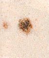
 Melanoma, brown and red lesion 1.jpg 562 × 712; 77 KB
-
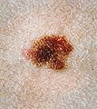
 Melanoma, brown lesion.jpg 562 × 712; 79 KB
-
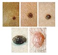
 Melanoma (1).jpg 684 × 1,026; 59 KB
-
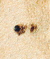
 Melanoma, red and brown lesion 2.jpg 562 × 712; 43 KB
-
 Squamous cell carcinoma (1).jpg 554 × 684; 70 KB
-
 Squamous cell carcinoma (2).jpg 1,025 × 683; 76 KB
-
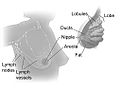 Breast anatomy.jpg 1,013 × 783; 62 KB
-
 Fink, diane.jpg 2,289 × 2,841; 660 KB
-
 Stethoscope and Laptop Computer - Nci-vol-9713-300.jpg 5,184 × 3,456; 9.16 MB
-
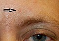 Trichilemmoma - Nci-vol-9808-72.jpg 373 × 259; 23 KB
-
 Metastatic Melanoma Cells Nci-vol-9872-300.jpg 3,000 × 3,000; 4.25 MB


.jpg)
_(1).jpg)

.jpg)

.jpg)

.jpg)
.jpg)
.jpg)
.jpg)
.jpg)
.jpg)
.jpg)


_(1).jpg)


_(1).jpg)


_(1).jpg)

_(1).jpg)

.jpg)
.jpg)


.jpg)

.jpg)


.jpg)
_-_crop.jpg)
.jpg)
.jpg)
.jpg)
.jpg)
.jpg)
.jpg)
.jpg)
_mastectomy_-_spanish.jpg)

.jpg)


.jpg)

.jpg)
.jpg)


