Category:Anatomy
Aller à la navigation
Aller à la recherche
Català: Anatomia
· Deutsch: Anatomie
· English: Anatomy
· Esperanto: Anatomio
· Français : Anatomie
· Íslenska: líffærafræði
· 日本語: 解剖学
· Português: Anatomia
· Tiếng Việt: Giải phẫu học
· Türkçe: Anatomi
· Walon : Antomeye
· 中文(简体):解剖学
· 中文(繁體):解剖學
· | Category Anatomy on sister projects: | |||||||||
|---|---|---|---|---|---|---|---|---|---|
ar: en: fr: gl: io: it: ja: ru: nl: no: pt: pl: th: zh: vi: |
Wiktionary | ||||||||
العربية: علم التشريح هو دراسة بنية جسم الإنسان (و الحيوان) و الكائنات الحيّة الأخرى مثل النباتات و الحشرات.
English: Anatomy is the study of the build of the human (and animal) body and other organisms such as plants and insects.
Español: La anatomía es el estudio de la estructura del cuerpo humano (y demás vertebrados), y de otros organismos (como las plantas y los insectos)
Français : L'anatomie est la science qui étudie les corps humains, animaux et végétaux
Galego: Anatomía é a ciencia que estuda a estrutura e forma dos seres orgánicos
Italiano: L'Anatomia è lo studio del corpo umano e degli altri organismi viventi.
Русский: Анатомия - наука о строении тела человека или животных, изученная на макроскопическом уровне. Представляет одну из ветвей Морфологии
Norsk bokmål: Anatomi er læren om byggningen av menneskers, samt dyr og andre organismers, kropp og struktur.
Polski: Anatomia to nauka zajmująca się budową ciała ludzi, zwierząt i innych organizmów żywych, takich jak rośliny.
ไทย: กายวิภาคศาสตร์เป็นวิชาซึ่งศึกษาเกี่ยวกับโครงสร้างของสิ่งมีชีวิต
Türkçe: Anatomi insan (ve hayvan) gövdesi ve bitkiler ile böcekler gibi diğer organizmaların yapısı üzerine çalışır.
Tiếng Việt: Giải phẫu học là môn học nghiên cứu hình thái và cấu tạo của cơ thể sinh vật.
Walon : L' antomeye, c' est l' syince ki discrît tot çou k' gn a dins l' coir d' ene djin.
中文:解剖学是研究生命体的结构和组织的一门科学。
Please use the following system when categorizing files:
- Place files relating to anatomy in this category. Anatomy is the scientific study of the structure of living things.
- Place files relating to bodies but not relating to their scientific study, such as ordinary images of parts of the body, in "Category:Body".
- Place files relating to animals in a suitable subcategory of "Category:Animal body".
- Place files relating to humans in a suitable subcategory of "Category:Human body".

discipline scientifique d'étude de la conformation animale (ou humaine)  | |||||
| Téléverser des médias | |||||
| Prononciation (fichier son) | |||||
|---|---|---|---|---|---|
| Nature de l’élément | |||||
| Sous-classe de | |||||
| Partie de | |||||
| Comprend | |||||
| À ne pas confondre avec | |||||
| |||||
Sous-catégories
Cette catégorie comprend 55 sous-catégories, dont les 55 ci-dessous.
*
A
- Acontia (anatomy) (1 F)
B
C
- Center for Anatomy (Charité) (11 F)
D
E
F
G
H
- Hilum (3 F)
I
L
M
- Manus (anatomie) (2 F)
O
- Organ size (19 F)
P
S
T
- Anatomy templates (3 P)
V
- Vinylite and corrosion (16 F)
Média dans la catégorie « Anatomy »
Cette catégorie comprend 388 fichiers, dont les 200 ci-dessous.
(page précédente) (page suivante)-
 024 Anatomy Laboratory.jpg 6 000 × 4 000 ; 5,61 Mio
-
 3D Interactive Anatomage Table .jpg 5 339 × 3 648 ; 1,52 Mio
-
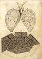
 Abeille, pétiole.jpg 2 687 × 2 257 ; 267 kio
-
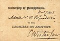 Admission ticket to Caspar Wistar lecture 1808.jpg 2 112 × 1 456 ; 977 kio
-
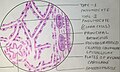
 Adénocytes buccaux de Wendilgarda (autre vue) et cuticule.jpg 1 795 × 1 143 ; 553 kio
-
 Adénocytes buccaux de Wendilgarda.jpg 1 218 × 2 003 ; 633 kio
-
 Agaricales Lame longueur et nuageuse.jpg 798 × 615 ; 66 kio
-
 Agaricales Lames insertion 3.jpg 1 080 × 546 ; 58 kio
-
 Agaricales Lames ventrues insertion 1.jpg 686 × 488 ; 50 kio
-
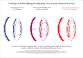
 Age-relation for aurofacial asymmetry.png 972 × 780 ; 127 kio
-
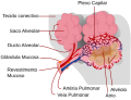 Alveolus diagram-pt.svg 373 × 283 ; 73 kio
-
 Anatomisches Institut Königsberg.jpg 977 × 563 ; 194 kio
-
 Anatomy class LCCN2014707319.tif 4 890 × 3 334 ; 15,55 Mio
-
 Anatomy Room. UMAR Puerto Escondido.png 1 920 × 1 080 ; 1,79 Mio
-
 Anatomy study.jpg 3 120 × 4 160 ; 2,55 Mio
-
 Anatomy.png 689 × 507 ; 24 kio
-
 AneurismaArteriaBasilaris 7361.jpg 5 184 × 3 456 ; 2,41 Mio
-
 ApiNATOMY conduit configurations.png 1 666 × 1 246 ; 1,04 Mio
-
 Arabic calligraphy by Anfal 3.jpg 1 920 × 1 090 ; 229 kio
-
 ArteryVeinCS.jpg 640 × 480 ; 187 kio
-
 Arthropod trachea.png 2 832 × 2 963 ; 7,82 Mio
-
 Artérias glúteas.png 892 × 482 ; 822 kio
-
 Baber-Contributions to the Minute Anatomy of the Thyroid Gland of the Dog.pdf 1 127 × 1 683, 14 pages ; 6,01 Mio
-
 Bayero university Anatomy department.jpg 4 368 × 2 016 ; 3,57 Mio
-
 Beheko oliba nukleoa.tif 1 602 × 916 ; 244 kio
-
 Bestibulu nukleoa isolatuta.jpg 166 × 117 ; 2 kio
-
 Bibliographie anatomique (1895) (20180034208).jpg 3 244 × 2 329 ; 1,07 Mio
-
 Blood Cooler Arm Data.png 1 096 × 460 ; 39 kio
-
 Bloqueio dos Canais de Sódio.png 304 × 379 ; 151 kio
-
 Cardiac muscle microscope.jpg 4 000 × 2 992 ; 3,84 Mio
-
 CardiacMuscle - longtitudinal.jpg 640 × 480 ; 188 kio
-
 Cellules, tissus, organes et systèmes.jpg 1 754 × 2 480 ; 842 kio
-
 Close-ups of various kinds of pathology affecting the tissues Wellcome L0074295.jpg 5 532 × 7 792 ; 13,96 Mio
-
 Comparative evolution of the striatum and pallium in vertebrates.png 2 902 × 1 370 ; 2,45 Mio
-
 Conducting system of the heart.png 2 550 × 3 350 ; 1,68 Mio
-
 Configurazione della tensostruttura animale (Barcelona 2008).jpg 3 008 × 2 000 ; 1,01 Mio
-
 Ctenophore diagram - en.svg 1 678 × 1 262 ; 165 kio
-
 Cuboidal Epithelium Section.jpg 640 × 480 ; 338 kio
-
 Cuticule buccale de Wendilgarda.jpg 1 133 × 1 833 ; 471 kio
-
 Cèl·lules del teixit ossi.jpg 1 064 × 747 ; 88 kio
-
 D1 (2).png 1 413 × 859 ; 344 kio
-
 Dana tapatuta.tif 1 286 × 872 ; 210 kio
-
 Deltoid muscle origin and insertion.jpg 920 × 1 043 ; 157 kio
-
 DenseConnectiveTissue-1.jpg 640 × 480 ; 184 kio
-
 DERICK 4444 (2).png 1 665 × 851 ; 441 kio
-
 DERICK 5.png 1 920 × 1 080 ; 786 kio
-
 DERICK 5555555.png 787 × 914 ; 456 kio
-
 DERICK.jpg 1 408 × 870 ; 123 kio
-
 Digitalizzato 20190102-1147-001.jpg 1 190 × 1 125 ; 531 kio
-
 Dinamite e seu Pavio.png 242 × 345 ; 48 kio
-
 Dissection of pregnant dogfish shark with exposed gills.jpg 807 × 1 076 ; 233 kio
-
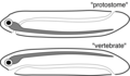 Dorsoventral inversion theory.png 2 997 × 1 750 ; 323 kio
-
 Dorsoventrale Inversionstheorie.png 3 037 × 1 750 ; 330 kio
-
 E. Geoffroy Saint-Hillaire, Philosophie anatomique. Wellcome L0029113.jpg 1 558 × 1 312 ; 1,01 Mio
-
 Effect of adrenalin on the cat bladder. Wellcome L0002006.jpg 1 795 × 997 ; 476 kio
-
 Electrical state of muscle fibres Wellcome L0001987.jpg 1 990 × 952 ; 801 kio
-
 Electrical state of muscle Wellcome L0001986.jpg 2 066 × 920 ; 572 kio
-
 Embryologie Zwerchfell.png 2 871 × 1 472 ; 682 kio
-
En-us-anatomy.ogg 1,2 s ; 14 kio
-
 Encyclopédie méthodique - Systeme anatomique, T01.djvu 4 267 × 6 400, 787 pages ; 91,99 Mio
-
 Encyclopédie méthodique - Systeme anatomique, T03 (page 9 crop).jpg 644 × 439 ; 80 kio
-
 Encyclopédie méthodique - Systeme anatomique, T03.djvu 2 339 × 2 985, 736 pages ; 38,75 Mio
-
 Encyclopédie méthodique - Systeme anatomique, T04.djvu 1 275 × 1 650, 575 pages ; 35,16 Mio
-
 Encyclopédie méthodique - Système anatomique, Planches.djvu 4 267 × 6 400, 261 pages ; 35,26 Mio
-
 Ensemble-des-corps.png 390 × 655 ; 184 kio
-
 Exercitationes Anatomicae de motu cordis Wellcome L0028762.jpg 1 054 × 1 967 ; 995 kio
-
 Experiments on animal electricity, 19th Century Wellcome L0001985.jpg 1 506 × 1 258 ; 624 kio
-
 Explication of female anatomical figure, 1680 Wellcome L0025084.jpg 1 080 × 1 790 ; 892 kio
-
 F. Glisson, Text to plate I,"Anatomia hepatis" Wellcome L0013984.jpg 1 105 × 1 717 ; 765 kio
-
 Fabricus Hildanus, Observationum et chirurgicarum... Wellcome L0026683.jpg 1 530 × 1 253 ; 812 kio
-
 Face-schema aurofacial asymmetry.png 324 × 363 ; 53 kio
-
 Fetal Pig Anatomy.png 1 053 × 802 ; 1,11 Mio
-
 Fibre net of Joseph von Gerlach Wellcome L0002013.jpg 907 × 2 047 ; 716 kio
-
 Fibrous skeleton of heart.png 2 550 × 3 350 ; 1,92 Mio
-
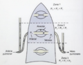 Fig 2. Zonas de West.png 880 × 689 ; 589 kio
-
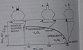 Fig 3. Diagrama.jpg 1 102 × 662 ; 59 kio
-
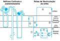 FIG. 6. 3. AMANDA (2).png 493 × 336 ; 169 kio
-
 FigOppositeAsymmetry.pdf 162 × 493 ; 9 kio
-
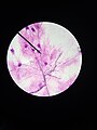
 Fingerprint under microscope.jpg 1 073 × 803 ; 168 kio
-
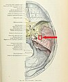 Foramen spinosum.jpg 582 × 700 ; 135 kio
-
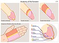 Foreskin Anatomy WIKI-EN.jpg 2 968 × 2 147 ; 1,29 Mio
-
 Frogdissectionsm.jpg 2 000 × 2 375 ; 3,9 Mio
-
 Front of forearm illustration. Wellcome L0012596.jpg 1 225 × 1 556 ; 839 kio
-
 Fundus part of stomach.jpg 2 288 × 1 065 ; 855 kio
-
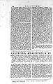 G. B. Morgagni, Epistolae anatomicae... Wellcome L0017155.jpg 1 066 × 1 680 ; 851 kio
-
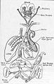
 Galen's physiological scheme Wellcome L0024393.jpg 1 162 × 1 612 ; 802 kio
-
 Galenask.jpg 257 × 399 ; 26 kio
-
 Gautier d'Agoty, muscles of eye, larynx.. Wellcome L0013458.jpg 1 222 × 1 546 ; 800 kio
-
 Gesch embryonal.png 671 × 900 ; 382 kio
-
 Glande rostrale de Diguetia.jpg 4 171 × 3 427 ; 892 kio
-
 Grant 1962 157.png 4 433 × 3 872 ; 31,52 Mio
-
 Grant 1962 157a.png 4 224 × 1 166 ; 11,39 Mio
-
 Grant 1962 157b.png 3 058 × 946 ; 6,51 Mio
-
 Greifswalder Anatomiegebäude.jpg 3 582 × 2 602 ; 1,7 Mio
-
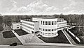
 GroningenAnatLab1938.jpg 750 × 1 053 ; 899 kio
-
 Gráfico de reabsorção da glicose.png 1 300 × 731 ; 62 kio
-
 H. Gray, Anatomy, descriptive..., 1897; the perinaeum Wellcome L0020687.jpg 1 232 × 1 524 ; 787 kio
-
 Halloween Horror Nights - American Horror Story (30465026281).jpg 1 824 × 2 736 ; 1,88 Mio
-
 Handcoloured woodcut Wellcome L0041481.jpg 2 660 × 3 664 ; 2,32 Mio
-
 Handcoloured woodcut Wellcome L0041482.jpg 2 668 × 3 680 ; 2,23 Mio
-
 Handcoloured woodcut Wellcome L0041483.jpg 2 672 × 3 680 ; 2,1 Mio
-
 Handcoloured woodcut Wellcome L0041484.jpg 2 672 × 3 636 ; 2,34 Mio
-
 Handcoloured woodcut Wellcome L0041485.jpg 2 668 × 3 708 ; 2,32 Mio
-
 Helicotrema 2.png 1 200 × 527 ; 130 kio
-
 Histology of Adipose tissue.jpg 2 472 × 1 445 ; 1,33 Mio
-
 Histology of cardiac muscle.jpg 2 403 × 1 467 ; 1,35 Mio
-
 Histology of Ileum.png 1 200 × 1 600 ; 1,91 Mio
-
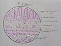 Histology of jejunum.jpg 3 000 × 2 244 ; 3,19 Mio
-
 Histology of keratinised epithelium.jpg 2 863 × 1 589 ; 1,73 Mio
-
 Histology of mixed salivary gland.jpg 1 200 × 1 600 ; 124 kio
-
 Histology of pseudo stratified ciliates columnar epithelium.jpg 2 326 × 1 367 ; 1,13 Mio
-
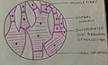
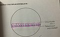 Histology of simple columnar epithelium.jpg 3 032 × 1 886 ; 1,99 Mio
-
 Histology of smooth muscle.jpg 2 488 × 1 447 ; 1,11 Mio
-
 Histology of stratified squamous non keratinised epithelium.jpg 2 283 × 1 365 ; 1,17 Mio
-
 Histology of transitional epithelium.jpg 2 577 × 1 402 ; 1,32 Mio
-
 Histology smooth muscle.jpg 2 879 × 1 640 ; 1,43 Mio
-
 Human body by rua.jpg 720 × 1 280 ; 156 kio
-
 Human Lens Scheimpflug layers.svg 1 488 × 1 052 ; 538 kio
-
 Human neuron.jpg 2 992 × 4 000 ; 2,95 Mio
-
 Human palatine bone (left).png 747 × 859 ; 312 kio
-
 Hydra Boby Wall Structure.jpg 2 844 × 1 584 ; 1,21 Mio
-
 Hymenoptera 1.3.png 2 146 × 1 585 ; 158 kio
-
Höla.ogg 0,7 s ; 11 kio
-
 Húmero humano.tif 2 480 × 3 508 ; 24,91 Mio
-
 III. bikote kranealeko nukleoa.png 596 × 495 ; 176 kio
-
 Illu conducting passages-pt-svg.svg 256 × 328 ; 21 kio
-
 Illustration of nervous system and nerve fibres Wellcome L0001861.jpg 1 192 × 1 614 ; 779 kio
-
 Illustration related to Vesalius. Wellcome M0016901.jpg 2 982 × 3 682 ; 4,59 Mio
-
 IMG Anatomy1.jpg 3 456 × 5 184 ; 5,84 Mio
-
 IMG Anatomy2.jpg 5 184 × 3 456 ; 5,37 Mio
-
 IMG Anatomy3.jpg 5 184 × 3 456 ; 4,96 Mio
-
 IMG Anatomy4.jpg 5 184 × 3 456 ; 5,61 Mio
-
 IMG Anatomy5.jpg 3 456 × 5 184 ; 5,01 Mio
-
 IMG Anatomy6.jpg 3 456 × 5 184 ; 5,66 Mio
-
 IMG Anatomy7.jpg 3 456 × 5 184 ; 5,73 Mio
-
 IMG Anatomy8.jpg 3 456 × 5 184 ; 5,26 Mio
-
 IMG Anatomy9.jpg 3 456 × 5 184 ; 5,44 Mio
-
 Inferior salivar nucleus.tif 1 048 × 603 ; 130 kio
-
 Inflammation of the capsule of the liver, with abscess. Wellcome L0033436.jpg 2 959 × 4 134 ; 2,69 Mio
-
 Insect ovariole diagram 2.svg 512 × 176 ; 4 kio
-
 Insect ovariole diagram.svg 512 × 1 018 ; 4 kio
-
Iraitz-aparatua.ogg 1 min 22 s ; 634 kio
-
 Items from the primitive medicine section Wellcome L0029857.jpg 1 298 × 1 535 ; 699 kio
-
 Ivory plaque showing a dissection scene in ebony frame, Euro Wellcome L0058568.jpg 4 152 × 2 832 ; 1,32 Mio
-
 J. Hutchinson, A smaller atlas of...clinical anatomy Wellcome L0027723.jpg 1 762 × 1 126 ; 1 013 kio
-
 J. Valverde de Hamusco, Anatomia del corpo humano... Wellcome L0030140.jpg 1 244 × 1 692 ; 1,14 Mio
-
 Jacob ben J. Soresina, Seder Ha-Nikhar Wellcome L0022143.jpg 1 635 × 1 133 ; 958 kio
-
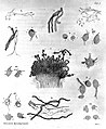
 Jardine Naturalist's library Entomology Plate I.jpg 1 782 × 2 856 ; 528 kio
-
 Jejunum.png 1 200 × 1 600 ; 1,98 Mio
-
Jer-Anatonmie.ogg 1,1 s ; 13 kio
-
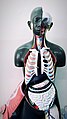
 Kidney anatomy model.jpg 1 280 × 720 ; 152 kio
-
 Kidneys of dog, 17th century Wellcome L0007974.jpg 1 014 × 1 828 ; 959 kio
-
 Krebsaugen, 2 pendants Wellcome M0016863.jpg 3 589 × 2 967 ; 1,25 Mio
-
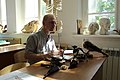
 Lab anato.jpg 800 × 600 ; 301 kio
-
 Lektion i anatomi vid Gymnastiska Centralinstitutet Stockholm kvinnliga kursen 1891-1893 gih0124.jpg 2 764 × 1 950 ; 3,65 Mio
-
 Liver illustration, in Specimen medicinae Sinicae. Wellcome L0010705.jpg 1 136 × 1 623 ; 438 kio
-
 Lobuli e lobi del cervelletto.jpg 3 140 × 1 673 ; 974 kio
-
 Lung histology.jpg 1 975 × 1 184 ; 950 kio
-
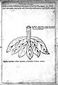 Lung illustration, in Specimen medicinae sinicae. Wellcome L0010701.jpg 1 124 × 1 646 ; 598 kio
-
 M. H. Dutrochet, Recherches anatomiques... Wellcome L0023902.jpg 1 108 × 1 806 ; 864 kio
-
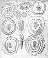 M. Malpighi, "Formatione pulli...", 1687 Wellcome L0000169.jpg 1 238 × 1 506 ; 829 kio
-
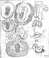 M. Malpighi, "Formatione pulli...", 1687 Wellcome L0000170.jpg 1 262 × 1 503 ; 824 kio
-
 Marcello Malpighi, De pulmonibus observation Wellcome L0031660.jpg 2 808 × 3 936 ; 4,09 Mio
-
 MOC fibers and the cholinergic synapse onto OHCs in the mature Organ of Corti.jpg 2 076 × 525 ; 111 kio
-
MP1 Pulmonology.webm 5 min 1 s, 1 280 × 720 ; 217,94 Mio
-
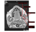 MRI showing masseter muscle and neighbors.png 933 × 835 ; 390 kio
-
 MS 564, Henri de Mondeville, Chirurgia... Wellcome L0032129.jpg 2 770 × 3 946 ; 4,38 Mio
-
 Muscles of the bank. Wellcome M0001605.jpg 1 368 × 1 793 ; 1,03 Mio
-
 Nakai Riken Anatomie.jpg 1 928 × 1 348 ; 537 kio
-
 Nerve cells and nerve fibres. Wellcome L0002196.jpg 1 276 × 1 558 ; 740 kio
-
 Nerve cells surrounded by axone terminations. Wellcome L0001463.jpg 2 042 × 930 ; 340 kio
-
 Nuck's canal in dog and human. Wellcome M0010704.jpg 4 000 × 2 665 ; 1,1 Mio
-
 Oesophagus.png 1 600 × 1 200 ; 2,04 Mio
-
 Opera omnia anatomica et chirurgica Wellcome L0060745.jpg 4 022 × 6 384 ; 6,14 Mio
-
 Opera omnia anatomica et chirurgica Wellcome L0060746.jpg 4 022 × 6 384 ; 5,74 Mio
-
 Ophthalmological instruments Wellcome L0002896.jpg 1 112 × 1 690 ; 966 kio
-
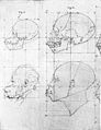 Optic Angle in chimpanzee and Negro, from De Lint. Wellcome M0000372.jpg 2 858 × 3 709 ; 5,27 Mio
-
 Oranda Geka Sho, 1840 Wellcome L0032565.jpg 3 543 × 5 315 ; 5,68 Mio
-
 Orchid aerial root anatomy showing the velamen.jpg 732 × 675 ; 757 kio
-
 Oseophagus histology of diagram.jpg 2 846 × 1 682 ; 1,51 Mio
-
 Ovarian disease Wellcome L0033435.jpg 2 976 × 4 110 ; 1,88 Mio
-
 Ovariole type diagram.svg 512 × 340 ; 12 kio
-
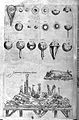
 Pancreas - apparaus used in de Graaf's experiments. Wellcome M0010451.jpg 2 632 × 4 128 ; 1,29 Mio
-
 Parts d'un os llarg.jpg 675 × 1 125 ; 69 kio
-
 Pectoralis Major.png 1 920 × 1 080 ; 735 kio
-
 Pencil anatomy of a nerve.jpg 2 304 × 3 456 ; 1,59 Mio
-
 Percent Italy.jpg 1 024 × 768 ; 81 kio
-
 Persistencia-es.jpg 304 × 261 ; 28 kio
-
 Persistencia.jpg 304 × 261 ; 33 kio
-
 Pharynx coronal section.jpg 1 280 × 960 ; 118 kio
-
 Pinax Mircocosmographicus. Wellcome L0010263.jpg 1 218 × 1 648 ; 864 kio
-
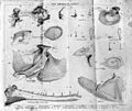
 Placenta, from H. Deventer "The art of midwifery improv'd." Wellcome L0020617.jpg 1 164 × 1 640 ; 1,1 Mio
-
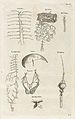
 Plate from Dutrochet, "Recherches Anatomiques," 1824 Wellcome L0033031.jpg 2 976 × 4 818 ; 5,44 Mio
-
 Plate from Haeckel, Anthropogenie Wellcome L0033034.jpg 3 414 × 5 436 ; 6,71 Mio
-
 Plate; "Memoire sur la Structure Elementaire...", Edwards Wellcome L0016384.jpg 1 250 × 1 520 ; 652 kio
-
 Plathelminthes Systeme de.svg 1 573 × 744 ; 461 kio
-
 Puente miodural 1.jpg 1 080 × 1 080 ; 336 kio
-
 Rajah 1.JPG 612 × 479 ; 43 kio
-
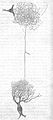
 Ramified nerve-cell, from grey matter. Wellcome M0011237.jpg 3 376 × 3 164 ; 4,59 Mio
-
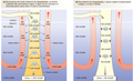
 Regiões do pulmão.png 819 × 629 ; 312 kio


_et_cuticule.jpg)


_(20180034208).jpg)


.jpg)


.png)


.png)


.jpg)


.png)


.jpg)


.png)


