Category:Zootomy
Jump to navigation
Jump to search
| Category Zootomy on sister projects: | |||||||||
|---|---|---|---|---|---|---|---|---|---|
Wiktionary |
Commons | ||||||||
anatomy of animals including humans | |||||
| Upload media | |||||
| Instance of | |||||
|---|---|---|---|---|---|
| Part of | |||||
| Said to be the same as | animal anatomy (Spanish Wikipedia) | ||||
| |||||
Subcategories
This category has the following 88 subcategories, out of 88 total.
- CT images of animals (35 F)
*
A
B
C
- Coelom (50 F)
- Cutaways of animals (3 F)
- Animal cuticles (12 F)
D
E
F
H
I
- Interstitial fluid (13 F)
J
K
L
M
N
- Nostrils in animals (1 F)
- Notochord (110 F)
O
- Oesophagus in animals (1 F)
P
- Photophores (12 F)
S
T
U
V
- Veins in animals (28 F)
- Vertebral column in animals (8 F)
W
Media in category "Zootomy"
The following 54 files are in this category, out of 54 total.
-
 A part of a uterus at the 9th month of gestation Wellcome L0033465.jpg 3,116 × 4,245; 3.17 MB
-
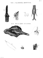
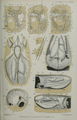
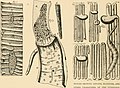
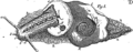
 A. Monro, "The structure...of fishes..." Wellcome L0028247.jpg 1,710 × 1,180; 1.1 MB
-
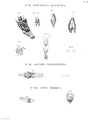
 A. Monro, "The structure...of fishes..." Wellcome L0028249.jpg 1,184 × 1,618; 843 KB
-
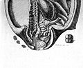 A. Monro, "The structure...of fishes..." Wellcome L0028250.jpg 1,542 × 1,270; 955 KB
-
 Aletaregeneracion.png 525 × 416; 64 KB
-
 Almohadillas.jpg 473 × 205; 19 KB
-
 Anatomy of a Physalia physalis colony.png 1,351 × 903; 210 KB
-
-
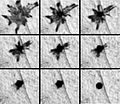
Autotomy.ogv 6.1 s, 320 × 240; 152 KB
-
 Captura de pantalla 2012-11-25 a las 17.01.58.png 675 × 208; 45 KB
-
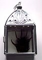 Curse reliquary.jpg 1,868 × 2,676; 367 KB
-
 DHM - Froschplatte.jpg 1,712 × 2,560; 2.15 MB
-
-
-
Dopamine-Modulates-Motor-Control-in-a-Specific-Plane-Related-to-Support-pone.0155058.s001.ogv 2 min 47 s, 640 × 480; 15.29 MB
-
-
-
Flapping-before-Flight-High-Resolution-Three-Dimensional-SkeletalKinematics-of-Wings-and-Legs-pone.0153446.s004.ogv 8.4 s, 1,440 × 1,080; 1.85 MB
-
-
 Foetus de veau.jpg 609 × 454; 53 KB
-
-
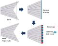
 Homocerca.png 1,439 × 1,079; 3.81 MB
-
 Iridiscent Meat.png 560 × 580; 318 KB
-
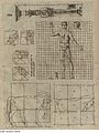
 Kuna Yala (2005) 14.jpg 706 × 1,058; 750 KB
-
 A. Monro, "The structure...of fishes..." Wellcome L0028248.jpg 1,264 × 1,622; 968 KB
-
 Larynx-Humboldt-Zoologie-T01p066.png 1,441 × 1,916; 383 KB
-
 Larynx-Humboldt-Zoologie-T02p068.png 1,434 × 1,910; 743 KB
-
 Larynx-Humboldt-Zoologie-T03p070.png 1,434 × 1,910; 485 KB
-
 Larynx-Humboldt-Zoologie-T04p072.png 1,437 × 1,910; 759 KB
-
 Melanophore.jpg 183 × 159; 29 KB
-
 Muybridge runner.jpg 424 × 318; 41 KB
-
 Neural.crest.cells.migration.svg 580 × 649; 50 KB
-
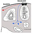 Neuralcrestroute.jpg 643 × 666; 260 KB
-
 Polet 6 organes limerotés.jpg 640 × 480; 75 KB
-
 PSM V76 D187 Graph of a frog sartorius muscle contractions.png 1,611 × 220; 72 KB
-
 PSM V76 D188 Graph of a frog gastrocnemius muscle contractions.png 822 × 592; 35 KB
-
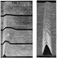 PSM V76 D189 Graph of a frog and rat gastrocnemius muscle contractions.png 1,617 × 1,638; 1 MB
-
 PSM V76 D190 Graph of a frog gastrocnemius muscle contractions.png 874 × 508; 48 KB
-
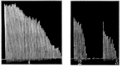 PSM V76 D194 Graph of a human finger flexor muscle contractions.png 1,613 × 887; 281 KB
-
 PSM V76 D197 Graph of a frog gastrocnemius muscle contractions.png 1,418 × 392; 109 KB
-
 PZSL1889Plate48.png 1,908 × 2,984; 5.85 MB
-
 PZSL1889Plate49.png 1,913 × 2,979; 5.66 MB
-
 Pánevní150.svg 744 × 1,052; 5 KB
-
 Rezets shlif.gif 258 × 86; 19 KB
-
 Skeletal sheep foot University of Dundee Museum collections.jpg 2,500 × 1,000; 902 KB
-
-
 Sprongbeen.jpg 700 × 469; 55 KB
-
 Sternopygus-aequilabiatus-Humboldt-Zoologie-T10p172.png 1,934 × 1,464; 702 KB
-
 Talus schaap CC BY NC ND 4 naturalis natuurwijzer.jpg 1,479 × 1,150; 444 KB
-
 The book of nature; or Fleuron T022987-10.png 1,115 × 440; 43 KB
-
 TheriaAnkle01.gif 320 × 285; 5 KB
-
 WalkTree.pdf 2,133 × 1,600; 705 KB
-
 Wikiproject Animal anatomy logo.svg 900 × 900; 264 KB
-
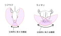 草食動物と肉食動物の視野.svg 500 × 300; 329 KB


_14.jpg)


_(14761488686).jpg)


