Category:Cell cultures
Naar navigatie springen
Naar zoeken springen
process by which cells are grown under controlled conditions | |||||
| Media uploaden | |||||
| Is een | |||||
|---|---|---|---|---|---|
| Subklasse van |
| ||||
| |||||
Ondercategorieën
Deze categorie bevat de volgende 7 subcategorieën, van de 7 in totaal.
C
- Contact inhibition (57 B)
H
- HEp-2 cells (4 B)
M
O
- Ovarian culture (6 B)
P
- Plaque-Assays (9 B)
R
- Rotary Cell Culture System (2 B)
Media in categorie "Cell cultures"
Deze categorie bevat de volgende 136 bestanden, van in totaal 136.
-
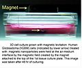 3D Cell Culturing by Magnetic Levitation Introduction Picture.jpg 395 × 326; 25 kB
-
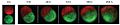 3D Cell Culturing by Magnetic Levitation Invasion Assay.jpg 974 × 204; 25 kB
-
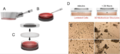
 3D Cell Culturing by Magnetic Levitation Protein Expression.jpg 973 × 273; 33 kB
-
-
 4T1 cell line.jpg 1.388 × 1.040; 298 kB
-
 6DIV 20X (3934022).jpg 851 × 1.182; 191 kB
-
 72hr sodyum48 saat.jpg 1.600 × 1.200; 272 kB
-
 96 well plate.jpg 5.881 × 3.921; 2,63 MB
-
-
-
-
-
-
-

-
 Alcian stain micromass.jpg 1.225 × 608; 544 kB
-
-
-
-
-
-
-
-
-
 Bacterial cell culture on agar plate ( DNA double helix conformation).jpg 2.974 × 2.847; 4,04 MB
-
 Bacterial cell culture on agar plate (Microscope conformation).jpg 4.000 × 4.292; 7,45 MB
-
 BallContamination.jpg 960 × 1.280; 84 kB
-
BioSig3D-High-Content-Screening-of-Three-Dimensional-Cell-Culture-Models-pone.0148379.s005.ogv 1 min 2 s, 976 × 720; 4,96 MB
-
BioSig3D-High-Content-Screening-of-Three-Dimensional-Cell-Culture-Models-pone.0148379.s006.ogv 3 min 39 s, 976 × 720; 13,5 MB
-
BioSig3D-High-Content-Screening-of-Three-Dimensional-Cell-Culture-Models-pone.0148379.s007.ogv 1 min 37 s, 964 × 720; 6 MB
-
BioSig3D-High-Content-Screening-of-Three-Dimensional-Cell-Culture-Models-pone.0148379.s008.ogv 2 min 12 s, 964 × 720; 10,87 MB
-
BioSig3D-High-Content-Screening-of-Three-Dimensional-Cell-Culture-Models-pone.0148379.s009.ogv 2 min 19 s, 964 × 720; 11,7 MB
-
BioSig3D-High-Content-Screening-of-Three-Dimensional-Cell-Culture-Models-pone.0148379.s010.ogv 3 min 19 s, 1.106 × 720; 21,04 MB
-
BioSig3D-High-Content-Screening-of-Three-Dimensional-Cell-Culture-Models-pone.0148379.s011.ogv 6,0 s, 318 × 333; 58 kB
-
BioSig3D-High-Content-Screening-of-Three-Dimensional-Cell-Culture-Models-pone.0148379.s012.ogv 6,2 s, 293 × 333; 51 kB
-
BioSig3D-High-Content-Screening-of-Three-Dimensional-Cell-Culture-Models-pone.0148379.s013.ogv 8,6 s, 320 × 333; 199 kB
-
BioSig3D-High-Content-Screening-of-Three-Dimensional-Cell-Culture-Models-pone.0148379.s014.ogv 8,0 s, 382 × 333; 354 kB
-
 Cell Culture (8145031511).jpg 889 × 895; 319 kB
-
 Cell culture-fig.png 1.152 × 1.059; 246 kB
-
 Cell on 2D Monolayer vs. 3D Hydrogel with RGD.png 1.320 × 291; 65 kB
-
 Cells in Cell Culture Dish.png 481 × 145; 11 kB
-
 Chk-UCH1-GFAP-20X-1.jpg 1.600 × 1.200; 3,07 MB
-
 CHO HeLa-myc DAPI DL20221106.png 4.181 × 1.537; 4,79 MB
-
 CO2 inkubaator rakukultuur DL20221105.jpg 4.608 × 3.456; 3,47 MB
-
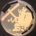
 Collezione Microbica.jpg 1.417 × 1.417; 1,33 MB
-
 CPCA-GFAP,MCA-5B10,Tau,neurons.jpg 1.000 × 1.000; 921 kB
-
 Culture de fibroblastes humains.tif 15.937 × 15.730; 230,62 MB
-
-
-
-
-
-
 E2P4 24 h ESC 10x DL20221106.png 3.647 × 1.281; 4,55 MB
-
 ELife-ZRK32-Fig5.jpg 1.736 × 1.812; 296 kB
-
 ELife-ZRK32-Fig5CD btm.jpg 766 × 595; 115 kB
-
-
 EMEM Sf9 rakukultuur DL20221105.jpg 4.608 × 3.456; 3,55 MB
-
 Epithelial-cells.jpg 202 × 202; 46 kB
-
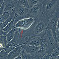
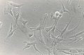
 Epithelioid cells and macrophages in cell culture.jpg 2.584 × 1.936; 3,8 MB
-
 Epithelioid cells of mice in the cell culture.jpg 1.318 × 875; 888 kB
-
 Fetal bovine serum.jpg 3.024 × 4.032; 2,48 MB
-
 HeLa Cell Culture Phase Contrast 2v1.png 2.880 × 1.440; 3,85 MB
-
 HeLa Cells Culture Phase Contrast 1 v1.jpg 1.920 × 1.440; 476 kB
-
 HeLa multipolar mitosis.jpg 1.370 × 1.371; 319 kB
-
 Human umbilical vein endothelial cells.jpg 2.659 × 2.618; 1,29 MB
-
 HUVECs.jpg 2.105 × 2.096; 1,08 MB
-
-
K63-Linked-Ubiquitination-Targets-Toxoplasma-gondii-for-Endo-lysosomal-Destruction-in-IFNγ-ppat.1006027.s001.ogv 3,4 s, 1.024 × 1.024; 307 kB
-
K63-Linked-Ubiquitination-Targets-Toxoplasma-gondii-for-Endo-lysosomal-Destruction-in-IFNγ-ppat.1006027.s002.ogv 2,8 s, 1.024 × 1.024; 134 kB
-
-
K63-Linked-Ubiquitination-Targets-Toxoplasma-gondii-for-Endo-lysosomal-Destruction-in-IFNγ-ppat.1006027.s005.ogv 3,3 s, 1.772 × 1.099; 1,85 MB
-
-
-
 Kluyveromyces lactis colony.png 144 × 121; 32 kB
-
 Kluyveromyces lactis plate.png 973 × 980; 1,14 MB
-
 Laminaar TYKI DL20221105.jpg 4.608 × 3.456; 3,51 MB
-
 Lonza-EBM2-Medium.JPG 680 × 1.024; 144 kB
-
LRRK2-Transport-Is-Regulated-by-Its-Novel-Interacting-Partner-Rab32-pone.0111632.s008.ogv 2,0 s, 2.136 × 1.024; 3,55 MB
-
LRRK2-Transport-Is-Regulated-by-Its-Novel-Interacting-Partner-Rab32-pone.0111632.s009.ogv 2,7 s, 864 × 434; 265 kB
-
 Mesothelioma Cells Culture.jpg 810 × 530; 34 kB
-
 Microglia Elisa Iba1 cultiu 3 placa 3B cd8.tif 1.376 × 2.066; 16,23 MB
-
-
-
-
-
 Ouroboros Steak.jpg 1.986 × 1.116; 548 kB
-
-
-
-
 Petri U2OS 20221105.jpg 3.456 × 4.608; 3,52 MB
-
 Phenol red pH 6,0 - 8,0.jpg 1.218 × 1.121; 56 kB
-
 Plaadid rakukultuur 20221105.jpg 4.608 × 3.456; 4,28 MB
-
Polarized-Cell-Division-of-Chlamydia-trachomatis-ppat.1005822.s004.ogv 16 s, 1.440 × 1.072; 125 kB
-
Polarized-Cell-Division-of-Chlamydia-trachomatis-ppat.1005822.s005.ogv 15 s, 1.440 × 1.072; 121 kB
-

-
 PubMed stats cell culture 3D O2 serum DL20221106.tif 2.736 × 2.568; 117 kB
-
 PubMed stats cell culture contam DL20221106.tif 2.700 × 2.394; 108 kB
-
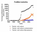
 PubMed stats cell culture DL20221106.tif 2.718 × 1.908; 78 kB
-
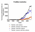
 PubMed stats cell culture primary line DL20221106.tif 2.718 × 2.502; 107 kB
-
 PubMed stats cell culture stem DL20221106.tif 2.718 × 2.550; 126 kB
-
 PubMed stats cell line culture DL20221106.tif 2.718 × 2.502; 114 kB
-
 Rakuloendur U87MG DL20221106.png 5.102 × 1.701; 16,46 MB
-
 Relda Cailleau 1965.tif 3.088 × 4.257; 12,54 MB
-
 Schéma de la micropropagation.png 1.280 × 720; 160 kB
-
 Scientist checks cell culture flask on microscope.jpg 1.000 × 1.250; 502 kB
-
 SF9 Cells phase contrast Lara Rudman.jpg 1.800 × 1.350; 459 kB
-
 Snu449 at 100 per cent confluency.jpg 1.600 × 1.200; 353 kB
-
 Snu449 at 50 to 60 per cent confluency.jpg 1.600 × 1.200; 292 kB
-
Spatial-Temporal-Study-of-Rab1b-Dynamics-and-Function-at-the-ER-Golgi-Interface-pone.0160838.s002.ogv 37 s, 1.920 × 1.080; 7,13 MB
-
-
Spatial-Temporal-Study-of-Rab1b-Dynamics-and-Function-at-the-ER-Golgi-Interface-pone.0160838.s004.ogv 50 s, 1.280 × 720; 4,59 MB
-
-
 STD Depth Coded Stack Slices through Cells.png 5.000 × 3.893; 22,96 MB
-
-
-
-
Targeting-MT1-MMP-as-an-ImmunoPET-Based-Strategy-for-Imaging-Gliomas-pone.0158634.s004.ogv 9,7 s, 736 × 736; 3,19 MB
-
 Test d'invasion de fibroblastes par des parasites Toxoplasma gondii.jpg 2.625 × 2.006; 549 kB
-
 Testis Organ Culture Vs Cell Culture.jpg 787 × 606; 76 kB
-
 Toxicology Research at FDA (NCTR 1242) (6009043802).jpg 3.987 × 2.648; 6,82 MB
-
 U251MG spheroid 24 h DL20221106.png 4.208 × 904; 2,24 MB
-
 Umbilical cord explants cell culture.jpg 2.048 × 1.536; 246 kB
-
Uveal-Melanoma-Cells-Utilize-a-Novel-Route-for-Transendothelial-Migration-pone.0115472.s001.ogv 15 s, 640 × 241; 8,12 MB
-
Uveal-Melanoma-Cells-Utilize-a-Novel-Route-for-Transendothelial-Migration-pone.0115472.s002.ogv 33 s, 717 × 177; 727 kB
-
Uveal-Melanoma-Cells-Utilize-a-Novel-Route-for-Transendothelial-Migration-pone.0115472.s003.ogv 11 s, 621 × 297; 9,34 MB
-
-
-
-
-
-
-
-
-
 Young Scientist.jpg 3.598 × 2.399; 1,48 MB
-
 Культура клітин Hela.jpg 3.396 × 2.547; 2,62 MB
-
 Культура стовбурових клітин нігтя.tif 1.600 × 1.200; 5,49 MB


.jpg)


.jpg)
.jpg)

.jpg)


_(6009043802).jpg)


