Category:Developmental biology
Jump to navigation
Jump to search

explores the processes by which individual organisms grow and develop (ontogeny)  | |||||
| Upload media | |||||
| Instance of | |||||
|---|---|---|---|---|---|
| Subclass of | |||||
| Has part(s) | |||||
| Different from | |||||
| |||||
Subcategories
This category has the following 60 subcategories, out of 60 total.
*
?
A
- ABC model (6 F)
B
- Developmental bias (8 F)
- Blastocoel (20 F)
- Blastocyst (51 F)
- The butterfly vivarium (8 F)
C
- Cell fate determination (154 F)
D
- Developmental signaling (78 F)
E
F
G
H
J
L
- Lithopedion (20 F)
M
- Media from BMC Developmental Biology (158 F)
- Media from Developmental Biology (16 F)
- Multicellularity (18 F)
- Myogenesis (4 F)
N
O
P
- Development of the pancreas (8 F)
- Pierre Pica (4 F)
R
S
- Somites (79 F)
T
V
Pages in category "Developmental biology"
This category contains only the following page.
Media in category "Developmental biology"
The following 200 files are in this category, out of 222 total.
(previous page) (next page)-
 3edg.png 1,674 × 893; 161 KB
-
 8-cell stage.jpg 1,224 × 1,632; 610 KB
-
 A human embryo of 2 mm. in median sagittal section.jpg 838 × 1,006; 310 KB
-
 A simplified representation of the essential steps in meisos.png 1,128 × 1,221; 424 KB
-

-
 Abb 9-4 Belyaev Experiment 21-10-10-B.jpg 1,942 × 1,183; 235 KB
-
 ABC Model with labels in Welsh.svg 512 × 391; 10 KB
-
 AcTub-Ath5.tif 600 × 600; 1.03 MB
-
 Age-relation for aurofacial asymmetry.png 972 × 780; 127 KB
-
 Anamorphic development in millipedes.tiff 1,843 × 3,926; 6.91 MB
-
 Arengubioloogia meetodid.png 480 × 640; 49 KB
-
 Asymmetric cell division neuroblast.jpg 236 × 301; 33 KB
-
 Asymmetric cell division.jpg 960 × 720; 61 KB
-
 Attracive-faces-symmetry.jpg 366 × 384; 18 KB
-
 Aurofacial asymmetry.pdf 695 × 385; 155 KB
-

-
 Axial twist in zebrafish embryo.pdf 1,204 × 866; 33 KB
-
 AxialTwistDevelopment.png 763 × 1,220; 479 KB
-
 AxialTwistScenario.png 642 × 310; 132 KB
-
 Bicoid (2).png 200 × 291; 4 KB
-
 Bicoid gradient photo.png 1,127 × 540; 184 KB
-
 Bicoid gradient.png 210 × 150; 7 KB
-
 Bicoid mutant.jpeg 850 × 763; 54 KB
-
 Bird song development timeline-sr.svg 617 × 500; 43 KB
-
 Bird song development timeline.png 617 × 500; 40 KB
-
 Bird song development timeline.svg 617 × 500; 43 KB
-
 BMP2.png 486 × 440; 43 KB
-
 BMP4 Signal Transduction Pathways.gif 720 × 540; 28 KB
-
 Bolachadapraia.jpg 3,264 × 2,448; 2.9 MB
-
 Bony nidus 1.jpg 395 × 237; 40 KB
-
 Branching morphogenesis in 3D cell culture.jpg 778 × 434; 98 KB
-
 CAM blood vessels.tif 1,377 × 2,074; 4.46 MB
-
 Cancer stem cells text resized-es.svg 930 × 690; 56 KB
-
 Cancer stem cells text resized.svg 930 × 690; 38 KB
-
 Cell Differentiation.jpg 727 × 384; 23 KB
-
 Cheryll Tickle Medal.jpg 7,236 × 3,672; 5.16 MB
-
 Clonal selection.svg 350 × 750; 137 KB
-
 Coelomate labeled.svg 250 × 250; 133 KB
-
 Coelomate.svg 250 × 250; 16 KB
-
 Comparison of cancer cell lines.png 640 × 285; 11 KB
-
 Comparison-of-Toll-pathways.png 814 × 581; 127 KB
-
 Complete cell lineage of C elegans.png 1,110 × 576; 41 KB
-
 Concs.gif 206 × 138; 632 KB
-
 Creode.jpg 265 × 190; 15 KB
-
-
-
-
-
-
-
 Development of embryonic nephrons.png 4,059 × 3,000; 1.8 MB
-
 Development of the retina.tif 6,560 × 1,312; 24.62 MB
-
 Diagrams showing the development of the amnion, chorion and allantois.jpg 1,269 × 1,151; 605 KB
-
 Dictyostelium Aggregation.JPG 973 × 768; 501 KB
-
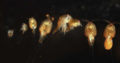
 Dictyostelium Fruiting Bodies.JPG 698 × 1,024; 204 KB
-
 Dictyostelium Late Aggregation 1.JPG 1,433 × 999; 2.86 MB
-
 Dictyostelium Myxamebas.JPG 1,215 × 866; 2.67 MB
-
 Dictyostelium Pseudoplasmodium.JPG 1,024 × 749; 516 KB
-
-
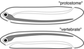 Dorsoventral inversion theory.png 2,997 × 1,750; 323 KB
-
 Dorsoventrale Inversionstheorie.png 3,037 × 1,750; 330 KB
-
 Dpp wing expression.jpg 216 × 324; 35 KB
-
Drosophila cleavage and gastrulation.webm 30 s, 1,920 × 800; 42.28 MB
-
 DrosophilaKutikula.jpg 1,181 × 591; 216 KB
-
 Duży inkubator.JPG 985 × 1,968; 250 KB
-
 Early embryonic development of Encarsia pergandiella. Nature as art or vice versa?.tif 4,164 × 3,120; 37.17 MB
-
 Eco-evo-devo-Fragen-klein2.jpg 1,856 × 1,180; 96 KB
-
 EctodermalSpecification.png 1,056 × 1,286; 96 KB
-
 EctodermalSpecification2.png 1,090 × 1,136; 158 KB
-
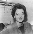 ElizabethDexterHay.png 1,114 × 1,138; 1.03 MB
-
 Embryonic development of the chondrocranium of a lizard.jpg 343 × 800; 140 KB
-
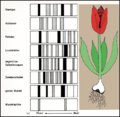 Enzymmmuster tulpe.png 700 × 682; 171 KB
-
 Equal vs unequal cleavage.jpg 720 × 540; 38 KB
-
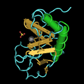
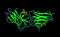 Estructura cristalina del dominio N-Reelin de F-spondin.png 858 × 535; 129 KB
-
 Even-skipped fushi tarazu.svg 203 × 121; 56 KB
-
 Experimente zur biologischen induktion.png 1,656 × 1,467; 298 KB
-
-
 Extensión disco imaginal de la pata.png 354 × 425; 23 KB
-
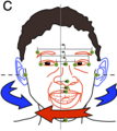 Face-schema aurofacial asymmetry.png 324 × 363; 53 KB
-
 Faringotrema.PNG 802 × 581; 19 KB
-
 Femaledevelopment.png 288 × 700; 42 KB
-
 Fetal hematopoieses.gif 600 × 595; 40 KB
-
 Fetale hematopoese.png 593 × 570; 27 KB
-
 FigOppositeAsymmetry.pdf 162 × 493; 9 KB
-
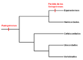
 Filogenia faringotremas.PNG 722 × 645; 138 KB
-
 Formation of the Primitive Streak.pdf 2,043 × 1,541; 14 KB
-
 Four diagrams showing hypothetical stages of early human embryos.jpg 1,631 × 1,434; 943 KB
-
 Gap ene expression-ja.png 200 × 205; 15 KB
-
 Gap gene expression.png 200 × 205; 11 KB
-
 Gap gene expression.svg 200 × 150; 5 KB
-
 Genes hox.jpeg 4,131 × 2,571; 857 KB
-
 Gill slits.png 753 × 645; 162 KB
-
 Grasshoppermetasnodgrass.jpg 529 × 932; 77 KB
-
 Gray34-ar.png 500 × 383; 66 KB
-
 Gray34.png 500 × 383; 36 KB
-
 HairyTeeth4TC.jpg 339 × 232; 35 KB
-
 Hatchlings by Sudiksha.jpg 2,272 × 1,704; 2.26 MB
-
 Heterochrony.svg 1,052 × 744; 24 KB
-
 High maintenance weed!.jpg 2,760 × 2,760; 2.78 MB
-
 Hiire embrüonaalsed tüvirakud diferentseerumas neuraalseteks eelasrakkudeks.JPG 2,560 × 1,920; 2.72 MB
-
 Homeotic selector gene complexes.svg 1,101 × 247; 56 KB
-
 How the Turtle Gets its Shell.svg 2,122 × 1,568; 54 KB
-
 Hox cluster.svg 419 × 129; 7 KB
-
 Human embryo Section of embryonic rudiment in Peters' ovum (first week).jpg 1,141 × 857; 540 KB
-
 Human embryonic stem cells.png 1,010 × 1,899; 2.64 MB
-
 Hypertrophic Zone of Epiphyseal Plate.jpg 408 × 574; 183 KB
-
 ICM signaling.jpg 960 × 720; 85 KB
-
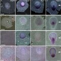
 Images representing technical steps during sEmbryo culture protocol.jpg 3,206 × 4,151; 1.98 MB
-
 Imaginal discs of drosophila.png 3,000 × 2,936; 869 KB
-
 Induktion var 3 007.jpg 2,434 × 1,701; 1.7 MB
-
 Insectwings2plain.svg 744 × 1,052; 110 KB
-
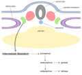 Intermediate mesoderm.png 486 × 447; 58 KB
-
 INVAGINATION.png 937 × 573; 19 KB
-
 Inver.jpg 1,024 × 768; 559 KB
-
 Invicta hatching.tif 1,024 × 768; 769 KB
-
 Ips cells ja.png 2,583 × 2,333; 373 KB
-
 Ips cells-es.png 2,022 × 1,928; 816 KB
-
 Ips cells.png 647 × 617; 69 KB
-
 Käfer004.jpg 1,200 × 1,704; 1.52 MB
-
 LarveKamsalamander.JPG 600 × 450; 205 KB
-
 Lateral inhibition process.jpg 906 × 468; 75 KB
-
 Lepisosteus oculatus larva at 22 days.png 440 × 361; 161 KB
-
 Lerva.jpeg 426 × 253; 43 KB
-
 Limb bud.png 850 × 496; 61 KB
-
 Lymphatic molecules.svg 881 × 819; 1,002 KB
-
 Macho con cuernos y sin cuernos de escarabajo O. taurus.png 969 × 872; 1,007 KB
-
 MAPKpathway diagram.svg 295 × 920; 33 KB
-
 MAPKpathway.png 294 × 920; 26 KB
-
 Mary Beth Hatten at Rockefeller University October 2, 2017.jpg 3,036 × 3,424; 6.24 MB
-
 Maternal effect mRNAs-2.svg 350 × 225; 2 KB
-
 Maternal-zygotic-transition.png 738 × 694; 59 KB
-
 MCR1erythro4TC.jpg 564 × 304; 41 KB
-
 Mechanism of neoblast specification during regeneration.jpg 3,529 × 2,647; 1.43 MB
-
 Mesenchymal Stem Cell.jpg 275 × 411; 60 KB
-
 Mesenchymal-Stem-Cell-rotate.jpg 411 × 275; 113 KB
-
 Monodelphis domestica embryo.png 303 × 222; 72 KB
-
 Morphogenetic.gif 279 × 267; 5 KB
-
 Morphogenic movements4567.jpg 720 × 540; 29 KB
-

-

-

-
 Mycoplasma genitalium.gif 280 × 280; 8 KB
-
 Má-formações Nêurula.png 355 × 434; 8 KB
-

-
 Neural crest.png 450 × 540; 39 KB
-
 Neurula.png 873 × 317; 21 KB
-
 Nodal signaling.jpg 1,168 × 1,198; 537 KB
-
 O.Hertwig1906Fig2-6.jpg 1,382 × 1,459; 347 KB
-
 Ontwikkelstadia wespenpoppen.jpg 681 × 1,979; 129 KB
-
 OPteloblast.jpg 958 × 1,508; 404 KB
-
 Organização corporal de Hydra.png 1,195 × 950; 178 KB
-
 Origen-Homeoticos.jpg 2,100 × 1,200; 102 KB
-
 Ovariole niche.png 680 × 496; 181 KB
-
 Ovum centrolecithale.png 600 × 600; 31 KB
-
 Ovum telolecithale.png 600 × 600; 41 KB
-
 P19 cell sorting out.png 228 × 223; 77 KB
-
 Pair rule.svg 336 × 334; 8 KB
-
 Phylogeny gill slit.png 782 × 546; 38 KB
-
 Physiological Polyspermy.png 1,360 × 604; 30 KB
-

-
 Pinus pinea foliage.jpg 762 × 850; 81 KB
-
 Polycheate Late Trochophore Lrva.jpg 5,069 × 6,572; 4.85 MB
-
 Polycheate Nereid Larva.jpg 4,318 × 5,292; 3.49 MB
-
-
-
-

-
 Primitiv Node.jpg 720 × 504; 42 KB
-
 Proneural genes in neurogenesis and gliogenesis pathway.png 2,650 × 1,095; 62 KB
-
 Prosobranchia- Late trochophore with protoconch.jpg 5,100 × 6,571; 4.75 MB
-
 Proteina-homeodominio.jpg 2,870 × 515; 195 KB
-
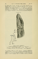
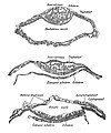
 PSM V84 D537 Facts and factors of development fig13.jpg 723 × 760; 111 KB
-
 PZSL1852PlateAnnulosa28.png 1,850 × 2,895; 3.45 MB
-
 PZSL1852PlateAnnulosa29.png 1,772 × 2,900; 3.98 MB
-
 PZSL1907Page054.png 2,079 × 3,213; 5.49 MB
-
 Raji cell lines.jpg 1,077 × 610; 138 KB
-
 RUH PMH.jpg 2,766 × 3,200; 1.33 MB
-
 Salivary gland epithelial tissue growing in vitro.jpg 542 × 540; 37 KB
-
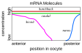
 Schematic diagram of the bicoid mRNA distribution.png 763 × 608; 26 KB
-

-
 Sea star regenerating legs.jpg 1,274 × 1,050; 529 KB
-
 Section showing three stages in the formation of the amnion of bat embryo.jpg 1,407 × 1,695; 911 KB
-

-
-
 Shark Lacerta deBeer1937.jpg 600 × 752; 178 KB
-
 Shh processing.png 791 × 307; 23 KB
-
 Shh processing.svg 790 × 305; 9 KB
-
 Shh structure.png 387 × 387; 70 KB
-
 Slack Essential Dev Biol Fig 02-08.jpg 255 × 482; 611 KB
-
 Slack Essential Dev Biol Fig 14.12a.jpg 1,146 × 913; 179 KB
-
 SMAD1.png 486 × 440; 68 KB
-
 SMAD2 SMAD4 COMPLEX.png 486 × 440; 110 KB
-
 SMAD3 SMAD4 COMPLEX.png 486 × 440; 110 KB
-
 SMURF2 SMAD7 complex.png 486 × 440; 87 KB
-
 SOFA-retina.tif 3,689 × 3,684; 38.88 MB
-

-

-
 Start-msc2 logo.png 588 × 106; 14 KB
-
 Stylopod-zygopod-autopod-pl.svg 397 × 131; 5 KB
-
 Stylopod-zygopod-autopod.png 397 × 201; 19 KB


.png)


.png)


.jpg)


_Development_-_Naturalist_Center_-_North_Carolina_Museum_of_Natural_Sciences.jpg)


_and_median_sagittal_sections_(g_and_h)_using_the_rabbit_as_a_model.jpg)


