Category:Chloroplasts
Vai alla navigazione
Vai alla ricerca
organello cellulare vegetale  | |||||
| Carica un file multimediale | |||||
| Istanza di |
| ||||
|---|---|---|---|---|---|
| Sottoclasse di | |||||
| |||||
Sottocategorie
Questa categoria contiene le 6 sottocategorie indicate di seguito, su un totale di 6.
File nella categoria "Chloroplasts"
Questa categoria contiene 120 file, indicati di seguito, su un totale di 120.
-
 03-11 Mnium3.jpg 1 730 × 1 206; 2,27 MB
-
 12 Cosmarium pseudamönum 48x25µm.jpg 436 × 346; 22 KB
-
 20090328 2222 Spirogyra.jpg 1 900 × 700; 106 KB
-
 20100417 005205 Zygnema.jpg 1 920 × 720; 118 KB
-
 20110331 012556 Algae.jpg 1 600 × 1 600; 252 KB
-
 Actinotaenium globosum 35x18µm.jpg 581 × 441; 32 KB
-
 Adenine Deaminates to Guanine vi.png 960 × 720; 93 KB
-
 Bryophyte Leaf Cells.jpg 2 982 × 1 870; 4,94 MB
-
 Calypogeia azurea Cells and blue oil bodies.jpg 3 200 × 2 400; 557 KB
-
 Carbon Concentrating Model for Chlamydomonas.png 960 × 720; 132 KB
-
 Chaperiony.png 794 × 1 123; 77 KB
-
-
-
 CHLOROPHYLL.jpg 2 448 × 3 264; 1,06 MB
-
 Chloroplast (Turkish).jpg 12 788 × 7 495; 6,78 MB
-
 Chloroplast 3.png 1 846 × 617; 1,57 MB
-
 Chloroplast and bacterial ribosome comparison.png 2 800 × 2 302; 3,27 MB
-
 Chloroplast de.png 815 × 586; 111 KB
-
 Chloroplast migration drill down diagram.png 1 201 × 765; 79 KB
-
 Chloroplast Nederlands.png 2 032 × 1 332; 1,12 MB
-
 Chloroplast pl.png 748 × 501; 314 KB
-
 Chloroplast Replication.png 1 280 × 1 024; 192 KB
-
 Chloroplast ribosome.jpg 600 × 598; 39 KB
-
 Chloroplast-drawing-lv.svg 1 297 × 746; 99 KB
-
 Chloroplast-drawing-numb.svg 1 033 × 685; 98 KB
-
 Chloroplast-drawing.svg 943 × 556; 87 KB
-
 Chloroplast-ger.png 815 × 586; 117 KB
-
 Chloroplast-japanese.jpg 748 × 501; 98 KB
-
 Chloroplast-new-Dutch.jpg 748 × 501; 66 KB
-
 Chloroplast-new.jpg 748 × 501; 54 KB
-
 Chloroplast.jpeg 1 383 × 1 390; 291 KB
-
 Chloroplast.png 744 × 438; 10 KB
-
 Chloroplaste-schema.png 509 × 324; 10 KB
-
 Chloroplasten.jpg 352 × 229; 31 KB
-
 Chloroplasto membranos.png 520 × 252; 12 KB
-
 Chloroplasts - Microscopic view of Elodea canadensis.jpg 2 592 × 1 944; 2,77 MB
-
 Chloroplasty.jpg 2 592 × 1 944; 439 KB
-
 Chromista structure.jpg 685 × 260; 105 KB
-
 Clorofila 3.jpg 1 632 × 1 224; 844 KB
-
 Cloroplast fases.gif 350 × 197; 8 KB
-
 Cloroplast.png 747 × 500; 83 KB
-
 Cloroplasti dentro cellule vegetali - Chloroplasts inside plant cells.jpg 2 484 × 2 336; 692 KB
-
 Cloroplasto.jpg 1 118 × 720; 101 KB
-
 Cloroplastos cel vegetal.jpg 845 × 454; 366 KB
-
-
-
 Cosmarium ochthodes vom 26.2.08 003bbc.jpg 196 × 227; 21 KB
-
 Cosmarium sp.jpeg 800 × 600; 56 KB
-
 Cosmarium.jpg 513 × 610; 174 KB
-
 CpDNA Replication vi.png 1 200 × 960; 156 KB
-
 Crs1 maize mutant.jpg 400 × 268; 80 KB
-
 CtDNA in circulation.png 726 × 520; 130 KB
-
 Drawing of Chloroplast.jpg 2 048 × 1 536; 573 KB
-
 Elodea chloroplasts 1 400×.png 1 700 × 1 700; 3,88 MB
-
 Elodea chloroplasts 100×.png 1 720 × 1 720; 5,83 MB
-
 Elodea chloroplasts 2 400×.png 1 680 × 1 680; 4,66 MB
-
 Elodea chloroplasts 3 400×.png 1 680 × 1 680; 4,49 MB
-
 Elodea chloroplasts 4 400×.png 1 691 × 1 690; 4,78 MB
-
 Elodea chloroplasts 5 400×.png 1 730 × 1 730; 5,29 MB
-
 Elodea chloroplasts 6 400×.png 1 680 × 1 680; 4,57 MB
-
 Elodea chloroplasts 7 400×.png 1 750 × 1 750; 5,01 MB
-
 Elodea-ciclose.gif 320 × 321; 3,04 MB
-
 Endosymbiotic theory.svg 512 × 384; 452 KB
-
 Esquema de la teoria de l'endosimbio siseriada.png 4 408 × 6 476; 2,96 MB
-
 Estructura del Cloroplasto.jpg 1 118 × 720; 125 KB
-
Euglena mutabilis.ogv 1 min 18 s, 720 × 480; 2,49 MB
-
 Features of a chloroplast (unannotated).jpg 600 × 400; 51 KB
-
 Features of a chloroplast.jpg 600 × 400; 73 KB
-
 Haeckel Desmidiea.jpg 2 332 × 3 267; 1,41 MB
-
-
-
-
Induction-of-protein-body-formation-in-plant-leaves-by-elastin-like-polypeptide-fusions-1741-7007-7-48-S1.ogv 5,0 s, 1 436 × 1 099; 780 KB
-
Induction-of-protein-body-formation-in-plant-leaves-by-elastin-like-polypeptide-fusions-1741-7007-7-48-S2.ogv 7,3 s, 1 013 × 1 099; 948 KB
-
Induction-of-protein-body-formation-in-plant-leaves-by-elastin-like-polypeptide-fusions-1741-7007-7-48-S3.ogv 4,7 s, 1 253 × 1 099; 792 KB
-
Induction-of-protein-body-formation-in-plant-leaves-by-elastin-like-polypeptide-fusions-1741-7007-7-48-S4.ogv 6,0 s, 1 106 × 1 099; 2,77 MB
-
-
Induction-of-protein-body-formation-in-plant-leaves-by-elastin-like-polypeptide-fusions-1741-7007-7-48-S6.ogv 13 s, 1 106 × 1 099; 2,62 MB
-
Induction-of-protein-body-formation-in-plant-leaves-by-elastin-like-polypeptide-fusions-1741-7007-7-48-S7.ogv 5,0 s, 1 106 × 1 099; 3,55 MB
-
Induction-of-protein-body-formation-in-plant-leaves-by-elastin-like-polypeptide-fusions-1741-7007-7-48-S8.ogv 5,0 s, 1 106 × 1 099; 1,36 MB
-
Induction-of-protein-body-formation-in-plant-leaves-by-elastin-like-polypeptide-fusions-1741-7007-7-48-S9.ogv 11 s, 1 106 × 1 099; 4,15 MB
-
-
 Kloroplastid.jpg 3 648 × 2 736; 1,15 MB
-
 Live leaf cells of the moss plant. మాస్ మొక్క పత్రం లోని హరిత రేణువుల తో కూడిన కణాలు.jpg 3 120 × 1 440; 2,1 MB
-
 Liverwort chloroplasts.jpg 1 919 × 1 279; 395 KB
-
Lophozia silvicola leaf cells.webm 18 s, 1 920 × 1 080; 11,46 MB
-
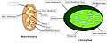 Mitochondria and Chloroplasts.jpg 1 302 × 516; 94 KB
-
-
-
 Mougeotia sp.jpeg 800 × 600; 39 KB
-
 NSRW Chlorophyll - Chloroplasts (1).jpg 457 × 400; 61 KB
-
 NSRW Chlorophyll - Chloroplasts (2).jpg 694 × 872; 179 KB
-
 Origin of mitochondria and chloroplasts.webp 3 235 × 1 442; 1,1 MB
-
-
 Plastid DNA scheme ru.png 800 × 566; 37 KB
-
Rapid-Mass-Movement-of-Chloroplasts-during-Segment-Formation-of-the-Calcifying-Siphonalean-Green-pone.0020841.s001.ogv 1 min 18 s, 640 × 480; 2,91 MB
-
 Red alga 100× objective oblique 1.jpg 4 592 × 3 056; 4,7 MB
-
 Red alga 100× objective oblique 2.jpg 4 592 × 3 056; 4,6 MB
-
 Red alga 100× objective.jpg 4 592 × 3 056; 4,07 MB
-

-
 S 24453197.jpg 628 × 657; 126 KB
-
 Scenedesmus quadricauda close up DIC Image-1.tif 828 × 973; 2,15 MB
-
Selective-Algicidal-Action-of-Peptides-against-Harmful-Algal-Bloom-Species-pone.0026733.s003.ogv 58 s, 680 × 512; 4,39 MB
-
Selective-Algicidal-Action-of-Peptides-against-Harmful-Algal-Bloom-Species-pone.0026733.s004.ogv 1 min 10 s, 680 × 512; 2,96 MB
-
 Spirogyra algae under microscope.jpg 2 048 × 1 536; 884 KB
-
 Spirogyra aumento de 1000x M.O. 02.JPG 4 000 × 3 000; 4,34 MB
-
 Spirogyra aumento de 1000x M.O. 04.JPG 4 000 × 3 000; 4,21 MB
-
 Spirogyra aumento de 400x M.O. 03.JPG 4 000 × 3 000; 4,24 MB
-
 Spirogyra cell.jpg 692 × 720; 82 KB
-
Structure-of-the-Chloroplast-Ribosome-Novel-Domains-for-Translation-Regulation-pbio.0050209.sv001.ogv 1 min 6 s, 426 × 320; 4,87 MB
-
 Tha4.jpg 375 × 247; 74 KB
-
 Thylakoid.png 960 × 720; 28 KB
-
 Thylakoid2.png 483 × 243; 18 KB
-
 Thylakoide.png 483 × 243; 23 KB
-
 Trad Chloroplast primary endosymbiosis.svg 500 × 320; 407 KB
-
 Überseemuseum Bremen 2009 238.JPG 4 272 × 2 848; 5,27 MB
-
 Клітини соснової голки.jpg 3 040 × 2 280; 3,44 MB
-
 Листик мха под микроскопом.jpg 3 440 × 2 580; 4,25 MB
-
 Ниткоподібні водорості під мікроскопом.jpg 4 148 × 3 088; 7,28 MB
-
 Хлоропласты 400.png 2 592 × 1 944; 6,44 MB


.jpg)


.jpg)


.jpg)
.jpg)

_leaf_protoplasts_observed_under_optical_microscopy.jpg)


