Category:Plant cells
Jump to navigation
Jump to search
eukaryotic cell present in green plants | |||||
| Upload media | |||||
| Instance of | |||||
|---|---|---|---|---|---|
| Subclass of |
| ||||
| Has part(s) | |||||
| |||||
Subcategories
This category has the following 26 subcategories, out of 26 total.
*
A
- Aerenchyma (18 F)
C
- Collenchyma (21 F)
M
N
P
- Plant mitochondria (5 F)
- Plasmodesmata (16 F)
- Preprophase band (9 F)
S
T
- Transfer cell (6 F)
V
W
Media in category "Plant cells"
The following 152 files are in this category, out of 152 total.
-
-
A-plausible-mechanism-for-auxin-patterning-along-the-developing-root-1752-0509-4-98-S6.ogv 22 s, 640 × 480; 478 KB
-
A-plausible-mechanism-for-auxin-patterning-along-the-developing-root-1752-0509-4-98-S9.ogv 23 s, 548 × 330; 227 KB
-
Allium cepa with KMnO4 and Phosphate.jpg 2,288 × 1,907; 816 KB
-
Allium-Differenzierung02-DM100x HF ba1.jpg 1,229 × 1,638; 1.73 MB
-
Allium-Differenzierung03-DM100x HF ba1.jpg 1,229 × 1,638; 1.42 MB
-
Allium-Differenzierung05-DM100x HF ba1.jpg 1,920 × 2,560; 1.62 MB
-
Amyloplasts.jpg 3,120 × 4,160; 2.42 MB
-
Anaphase of plant cell mitosis.jpg 894 × 648; 36 KB
-
Aufbau des Blattes.jpg 3,508 × 2,480; 1.32 MB
-
Aulacomnium palustre lamina.jpeg 800 × 600; 121 KB
-
Barbula unguiculata lamina.jpeg 800 × 600; 107 KB
-
Bartramia ithyphylla blattscheide.jpeg 800 × 600; 102 KB
-
Brachythecium velutinum lamina.jpeg 800 × 600; 67 KB
-
Bryophyte Leaf Cells.jpg 2,982 × 1,870; 4.94 MB
-
Bryum caespiticium lamina.jpg 800 × 600; 102 KB
-
Bryum capillare lamina.jpeg 800 × 600; 98 KB
-
Bryum elegans lamina.jpeg 800 × 600; 101 KB
-
Bryum elegans rhizoide.jpeg 800 × 600; 73 KB
-
Bryum imbricatum blattgrund.jpeg 800 × 600; 113 KB
-
Bryum imbricatum lamina.jpeg 800 × 600; 85 KB
-
Bryum turbinatum lamina.jpeg 800 × 600; 142 KB
-
Cell membrane in Rheo leaf cell.jpg 3,120 × 1,440; 1.71 MB
-
Cells in an onion root under light microscope.jpg 3,072 × 4,080; 2.68 MB
-
Cells in Leaf.jpg 5,164 × 2,505; 3.34 MB
-
Cephalozia connivens flankenblatt.jpeg 800 × 600; 58 KB
-
Clorofila 3.jpg 1,632 × 1,224; 844 KB
-
Corte longitudinal arveja.JPG 3,648 × 2,736; 740 KB
-
Corte Phyrus communis.JPG 3,648 × 2,736; 666 KB
-
Cromoplastos tomate.jpg 704 × 986; 99 KB
-
Cyclose.ogv 13 s, 1,280 × 1,024; 1.32 MB
-
Cyperus alternifolius, stalk, Etzold green 9.jpg 1,911 × 1,257; 618 KB
-
Células vegetais vistas sob um microscópio de luz.jpg 2,448 × 3,264; 4.25 MB
-
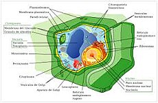
Differences between simple animal and plant cells (en).svg 512 × 253; 7 KB
-
Differences between simple animal and plant cells (numbers).svg 983 × 539; 62 KB
-
-
-
-
Drepanocladus polycarpus blattfluegel.jpeg 800 × 600; 118 KB
-
Drepanocladus polycarpus lamina.jpeg 800 × 600; 106 KB
-
EB1911 Plants - examples of cell differentiation (2).jpg 726 × 586; 123 KB
-
EB1911 Plants - examples of cell differentiation.jpg 722 × 405; 74 KB
-
-
-
Elodea cells under microscope.jpg 3,024 × 4,032; 3.04 MB
-
Epidermal Cells of Plant Leaf..jpg 2,048 × 1,887; 2.09 MB
-
Epidermis of Festuca arundinacea.jpg 8,499 × 6,178; 9.12 MB
-
Epitelio cebolla 2.jpg 755 × 773; 391 KB
-
Esquema de la teoria de l'endosimbio siseriada.png 4,408 × 6,476; 2.96 MB
-
Ficusxylem.jpg 1,771 × 991; 304 KB
-
Figure 04 03 01b.png 544 × 459; 108 KB
-
Foto wiki.jpg 800 × 585; 152 KB
-
Green algae .jpg 1,215 × 795; 190 KB
-
Hypnum cupressiforme perichaetialblaetter.jpeg 800 × 600; 75 KB
-
Inside the peppers.jpg 3,631 × 2,462; 3.02 MB
-
JZn84a7-hSE.jpg 719 × 960; 123 KB
-
Komórki Symphoricarpos Duhamel, Cell.jpg 950 × 713; 676 KB
-
Leaf palisade mesophyll.jpg 483 × 640; 271 KB
-
Leaf spongy mesophyll.jpg 480 × 640; 310 KB
-
Lepidozia reptans zellen.jpeg 800 × 600; 55 KB
-
-
-
-
-
-
-
Live leaf cells of the moss plant. మాస్ మొక్క పత్రం లోని హరిత రేణువుల తో కూడిన కణాలు.jpg 3,120 × 1,440; 2.1 MB
-
Längsschnitt Mark Große Kapuzinerkresse.JPG 3,648 × 2,736; 2.36 MB
-
Meyers b2 s0435 b1.png 262 × 541; 31 KB
-
Mitochondria-Change-Dynamics-and-Morphology-during-Grapevine-Leaf-Senescence-pone.0102012.s002.ogv 4.3 s, 512 × 512; 583 KB
-
Mitochondria-Change-Dynamics-and-Morphology-during-Grapevine-Leaf-Senescence-pone.0102012.s003.ogv 4.3 s, 512 × 512; 582 KB
-
Mitochondria-Change-Dynamics-and-Morphology-during-Grapevine-Leaf-Senescence-pone.0102012.s004.ogv 4.3 s, 1,024 × 1,024; 4.3 MB
-
Mitochondria-Change-Dynamics-and-Morphology-during-Grapevine-Leaf-Senescence-pone.0102012.s005.ogv 4.3 s, 1,024 × 1,024; 5.27 MB
-
Moss chloroplasts 100× objective oblique.jpg 2,200 × 1,200; 882 KB
-
Moss chloroplasts 100× objective.jpg 2,400 × 1,250; 998 KB
-
Onion skin under microscope.jpg 2,592 × 1,944; 1.92 MB
-
Overlapping TIP isoforms are mainly detected at the tonoplast of the central vacuole.jpg 1,200 × 2,180; 685 KB
-
Paludella squarrosa blattspitze.jpeg 800 × 600; 107 KB
-
Parastās priedes (Pinus sylvestris) koksnes tangenciālā griezuma attēls.jpg 3,456 × 2,304; 1.61 MB
-
Parastās priedes (Pinus sylvestris) koksnes šķērsgriezuma attēls.jpg 3,263 × 2,447; 2.37 MB
-
Pellia endiviifolia thallus.jpeg 800 × 600; 94 KB
-
Pellia endiviifolia thallusrand.jpeg 800 × 600; 108 KB
-
Penampang membujur daun Rhoeo discolor.png 268 × 472; 241 KB
-
Physcomitrium pyriforme lamina.jpeg 800 × 600; 109 KB
-
Plagiomnium elatum blattsaum.jpeg 800 × 600; 141 KB
-
Plagiomnium ellipticum lamina.jpeg 800 × 600; 142 KB
-
Plant cell structure.jpg 1,280 × 720; 98 KB
-
Plant cell under microscope.jpg 2,445 × 1,989; 1.07 MB
-
Plant Cells, Being Plasmolyzed.jpg 960 × 1,149; 207 KB
-
Plant tissue from a colon biopsy specimen (5297666726).jpg 1,383 × 1,382; 998 KB
-
-
Plant-stem-cell-maintenance-involves-direct-transcriptional-repression-of-differentiation-program-msb20138-s11.ogv 1 min 11 s, 1,080 × 360; 13.37 MB
-
Plasmolysis&deplasmolysis.jpg 2,448 × 3,264; 1.62 MB
-
Plazmolyzed Elodea Cells under 400X Magnification.jpg 1,708 × 1,707; 928 KB
-
Pleurotaenium Species of Desmid.jpg 2,982 × 1,984; 3.46 MB
-
Pohlia melanodon lamina.jpeg 800 × 600; 88 KB
-
Pohlia nutans lamina.jpeg 800 × 600; 105 KB
-
Potato starch.jpg 600 × 483; 82 KB
-
Prašník a pyl Salvia sp..jpg 4,080 × 3,072; 4.78 MB
-
Ptilidium ciliare laminazellen.jpeg 800 × 600; 74 KB
-
PumpkinStemcs400x2.jpg 1,024 × 768; 154 KB
-
PumpkinStemcs400x4.jpg 1,024 × 768; 148 KB
-
PumpkinStemcs400x9.jpg 1,024 × 768; 167 KB
-
-
-
-
-
Rasprostranjenje hloroplasta.PNG 426 × 325; 9 KB
-
Red onion cells (microscope observation).jpg 3,120 × 4,160; 2.06 MB
-
Red onion cells.jpg 2,988 × 5,312; 1.87 MB
-
Research SCPL-2.jpg 714 × 536; 63 KB
-
Rhoeo Discolor - Plasmolysis.jpg 1,656 × 1,527; 810 KB
-
Rhoeo Discolor epidermis.jpg 795 × 729; 209 KB
-
Riccardia chamedryfolia querschnitt.jpeg 800 × 600; 73 KB
-
Riccardia chamedryfolia zellen.jpeg 800 × 600; 99 KB
-
RiceStemcs400x5.jpg 1,024 × 768; 162 KB
-
RiceStemcs600x3.jpg 1,024 × 768; 105 KB
-
RiceStemcs600x5.jpg 1,024 × 768; 111 KB
-
RiceStemcs600x6.jpg 1,024 × 768; 113 KB
-
Roma Tomato Skin 600x.jpg 640 × 480; 196 KB
-
Scharbockskrautknolle Querschnitt differenziertes Speichergewebe.JPG 2,816 × 2,112; 2.28 MB
-
SeminalBudStemTipls400x1.jpg 1,024 × 768; 215 KB
-
SeminalBudStemTipls400x3.jpg 1,024 × 768; 212 KB
-
Slupka plodu Tamarindus indica.jpg 4,080 × 3,072; 3.13 MB
-
Slupka Tamarindus indica.jpg 4,080 × 3,072; 4.36 MB
-
Stem-Collenchyma400x1.jpg 1,024 × 768; 166 KB
-
Stem-Collenchyma400x3.jpg 1,024 × 768; 178 KB
-
Stem-Sclerenchvma400x2.jpg 1,024 × 768; 181 KB
-
Stoma in an onion.jpg 2,448 × 3,264; 414 KB
-
Tamaños.png 524 × 382; 24 KB
-
The Annals and magazine of natural history (1841) (17790786383).jpg 2,506 × 4,123; 874 KB
-
-
-
-
-
Tomatillo Husk 150x.jpg 640 × 480; 220 KB
-
Trad2.jpg 1,772 × 1,168; 1.69 MB
-
Trad3.jpg 1,776 × 1,168; 868 KB
-
Trad4.jpg 1,776 × 1,168; 650 KB
-
Turgid Elodea Cells under 400X Magnification.jpg 1,705 × 1,705; 996 KB
-
Vacuola vegetal.png 928 × 570; 175 KB
-
VesiclesPlantsc.jpg 702 × 362; 87 KB
-
-
Õuna koore pigmente sisaldavad rakud.jpg 2,023 × 1,505; 2.94 MB
-
Ευκαρυωτικό κύτταρο (φυτικό).png 982 × 678; 481 KB
-
Плазмолиз клетки кожицы лука.jpg 3,120 × 4,160; 2.65 MB
-
Поперечный срез стебля кубышки малой.jpg 1,155 × 2,048; 194 KB
-
Спираль жизни.tiff 2,776 × 2,080; 16.54 MB
-
表皮細胞-2.jpg 1,227 × 267; 104 KB
-
表皮細胞.tif 236 × 199; 80 KB


