Category:Cysts
Zur Navigation springen
Zur Suche springen
Radiologie: Ultraschall · Computertomographie · Magnetic resonance · Positronen-Emissions-Tomographie | Anatomische Pathologie: Gross pathology · Histopathologie | Othering: Bauchspiegelung | Dateiformat: Scalable Vector Graphics | 
von einem epithelialen Häutchen ausgekleideter Hohlraum im Gewebe  | |||||
| Medium hochladen | |||||
| Ist ein(e) | |||||
|---|---|---|---|---|---|
| Unterklasse von | |||||
| |||||
Unterkategorien
Es werden 51 von insgesamt 51 Unterkategorien in dieser Kategorie angezeigt:
In Klammern die Anzahl der enthaltenen Kategorien (K), Seiten (S), Dateien (D)
B
- Bartholin's cyst (8 D)
C
- Choledochal cysts (2 D)
- Coccygeal cysts (2 D)
- Cystic acne (3 D)
D
E
- Epididymal cysts (6 D)
- Eye cysts (2 D)
G
J
L
M
N
O
P
- Preauricular sinus and cyst (5 D)
R
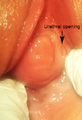
- Ranula (3 D)
- Rathke's cleft cyst (1 D)
S
- Splenic cysts (6 D)
T
V
- Vocal fold cyst (2 D)
Medien in der Kategorie „Cysts“
Folgende 52 Dateien sind in dieser Kategorie, von 52 insgesamt.
-
 2016-11-27 19-29-43 nodule.jpg 3.648 × 2.432; 3,23 MB
-
 2016-11-27 19-29-56 nodule.jpg 3.648 × 2.736; 3,3 MB
-
 2016-11-27 19-30-18 nodule.jpg 3.648 × 2.736; 2,59 MB
-
 2016-11-27 19-30-57 nodule.jpg 3.648 × 2.736; 3,24 MB
-
 2016-11-27 19-31-32 nodule.jpg 1.824 × 2.736; 2,18 MB
-
 2016-11-28 08-44-03 nodule.jpg 3.648 × 2.736; 2,52 MB
-
 2016-11-28 13-44-54 nodule.jpg 3.398 × 2.423; 2,37 MB
-
 2016-11-28 14-06-06 nodule.jpg 2.374 × 1.425; 951 KB
-
 2016-11-28 14-54-06 nodule.jpg 1.981 × 2.736; 1,75 MB
-
 2023-07-18 11-09-33 loupe-crane.jpg 4.000 × 3.000; 7,32 MB
-
 2023-07-18 11-10-22 loupe-crane.jpg 4.000 × 3.000; 7,86 MB
-
 2023-07-18 17-52-28 loupe-crane.jpg 1.379 × 1.511; 2,11 MB
-
 2023-07-19 08-56-46 kyste.jpg 4.000 × 3.000; 6,76 MB
-
 2023-07-19 08-56-54 kyste.jpg 1.308 × 979; 740 KB
-
 2023-07-19 08-57-02 kyste.jpg 4.000 × 3.000; 6,17 MB
-
 2023-07-19 09-02-30 kyste.jpg 3.717 × 2.787; 7,26 MB
-
 American quarterly of roentgenology (1909) (14757093422).jpg 2.024 × 2.022; 1,3 MB
-
 American quarterly of roentgenology (1909) (14757406945).jpg 2.286 × 2.286; 1,78 MB
-
 Annual of the universal medical sciences (1895) (14598134419).jpg 1.360 × 858; 422 KB
-
 Cystic adenoma removed from the left lobe of a thyroid gland Wellcome L0061704.jpg 5.118 × 4.746; 3,56 MB
-
 Diseases of the heart and thoracic aorta (1884) (14784899595).jpg 2.053 × 3.502; 896 KB
-
 EB1911 Gregarines - Cyst of Gregarina blattarum.jpg 494 × 419; 52 KB
-
 Egg, Cyst and Worms Preservatives.jpg 2.048 × 1.536; 813 KB
-
 Entamoeba coli cysts.jpg 448 × 327; 95 KB
-
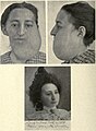 Epithelioma and Cyst.jpg 1.618 × 2.206; 673 KB
-
 Giardia cyst.JPG 1.192 × 803; 63 KB
-
 Glioependymal cyst HE.jpg 2.080 × 1.542; 950 KB
-
 Gould Pyle 235.jpg 720 × 979; 261 KB
-
 Gynecological diagnosis (1910) (14775039741).jpg 736 × 1.256; 275 KB
-
 Gynecology (1916) (14592559407).jpg 1.596 × 1.596; 572 KB
-
 Kystes méningés images retardées.jpg 480 × 360; 33 KB
-
 LargeAbdoCyst2008.jpg 3.072 × 2.304; 604 KB
-
 Operative gynecology - (1906) (14596888839).jpg 1.218 × 876; 187 KB
-
 Operative gynecology - (1906) (14760517636).jpg 1.608 × 2.516; 1,16 MB
-
 Paraprostatic Cyst Dog.jpg 356 × 348; 28 KB
-

-

-

-
 Parasite140085-fig3 Echinococcus granulosus cysts from boiled livers and lungs of sheep.tif 2.039 × 2.998; 4,78 MB
-
 Parasite140120-fig4 Acanthamoeba keratitis.png 5.950 × 1.985; 2,2 MB
-
 Parasite180022-fig1 Cystic echinococcosis in a cat.png 2.120 × 742; 511 KB
-
 Pathology and treatment of diseases of women (1912) (14595000168).jpg 1.260 × 628; 170 KB
-
 PCOS.jpg 289 × 407; 76 KB
-
 Rat Mammary Cyst.JPG 430 × 271; 17 KB
-
 Resting cysts of dinoflagellates.webp 3.802 × 1.904; 2,74 MB
-
 Skene's duct cyst-ar.png 309 × 451; 298 KB
-
 Skene's duct cyst.png 309 × 451; 171 KB
-
 The diagnosis and treatment of diseases of women (1907) (14597798870).jpg 998 × 1.482; 386 KB
-
 The diagnosis of diseases of women (1905) (14763150072).jpg 1.922 × 1.600; 439 KB
-
 The pathology and surgical treatment of tumors (1895) (14796908053).jpg 732 × 1.260; 325 KB
-
 Thyroglossal cyst.jpg 3.264 × 1.836; 1,77 MB
-
 Traumatisch rupturierte Nierenzyste - CT axial und coronar - 001.jpg 2.672 × 1.098; 256 KB


_(14757093422).jpg)
_(14757406945).jpg)
_(14598134419).jpg)

_(14784899595).jpg)


_(14775039741).jpg)
_(14592559407).jpg)


_(14596888839).jpg)
_(14760517636).jpg)


_(14595000168).jpg)


_(14597798870).jpg)
_(14763150072).jpg)
_(14796908053).jpg)
