Category:Tumors
Jump to navigation
Jump to search
Radiology: Ultrasound · X-ray · Computed tomography · Magnetic resonance · Positron emission tomography · Bone scintigraphy | Anatomical pathology: Cytopathology · Gross pathology · Histopathology | Other: Endoscopy · Laparoscopy · Epidemiology | File format: SVG · Video recording | 

![]() Not redirect this category to Category:Neoplasms. Thus Category:Cysts are tumors but not neoplasms.
Not redirect this category to Category:Neoplasms. Thus Category:Cysts are tumors but not neoplasms.
abnormal growth of tissue forming a mass  | |||||
| Upload media | |||||
| Subclass of | |||||
|---|---|---|---|---|---|
| Different from | |||||
| |||||
Subcategories
This category has the following 36 subcategories, out of 36 total.
Media in category "Tumors"
The following 46 files are in this category, out of 46 total.
-
 3D still showing tumor.jpg 1,280 × 720; 562 KB
-
 A3A.jpg 216 × 378; 74 KB
-
 Ajna tumor.jpg 1,080 × 980; 114 KB
-

-
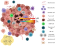 Components-of-the-tumor-microenvironment.png 850 × 680; 155 KB
-
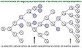 DarwinCancer.jpg 900 × 530; 50 KB
-
 Detail of secondary cancerous tumour swelling in the head of Wellcome V0036278EBL.jpg 1,600 × 1,180; 952 KB
-
 Dysembryoplastic neuroepithelial tumor.jpg 614 × 571; 79 KB
-
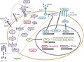
 Esquema de les interaccions establertes per LIGHT i consequencies.jpg 1,152 × 720; 89 KB
-
 Esquema que mostra les consecuències de la unió de DcR3 a LIGHT.png 598 × 319; 32 KB
-
 Excision of a tumour causing epilepsy, 1886. Wellcome M0016258.jpg 2,563 × 4,114; 1.86 MB
-
 Faceted Search example.jpg 1,920 × 1,160; 458 KB
-
 Fatty tumor.jpg 1,241 × 1,503; 860 KB
-
 GIST case report.pdf 1,275 × 1,650, 3 pages; 1.27 MB
-
 Gross specimen of liver angiosarcoma.jpg 844 × 634; 223 KB
-
 Human colon tumor.png 556 × 521; 459 KB
-
 Interaccions establertes per LIGHT.jpg 2,338 × 1,700; 281 KB
-
 Malignant growth springing from the eye of a woman Wellcome L0061858.jpg 4,552 × 5,224; 4.16 MB
-
 Meningioma of the sagittal sinus isolated.jpg 2,000 × 1,146; 396 KB
-
 Meningioma of the sagittal sinus.jpg 2,000 × 1,152; 592 KB
-
 MMHCdb Logo.jpg 640 × 215; 35 KB
-
 Multilocular Hydatid Tumor.jpg 1,228 × 1,541; 889 KB
-
 NIR heptamethine dye on a tumor.png 483 × 248; 54 KB
-
 Patiënt met een rugontsteking in het ziekenhuis van Penang, Bestanddeelnr 255-6745.jpg 2,572 × 2,580; 500 KB
-
 PDX Like Me.jpg 1,920 × 1,160; 409 KB
-
 Rhabdoidtumourcell.jpg 128 × 144; 38 KB
-
 Secondary cancerous tumour swelling in the head of a 49-year Wellcome V0036278ETL.jpg 1,560 × 1,352; 1.06 MB
-
 Section of a boy's head with a tumour Wellcome L0061565.jpg 4,673 × 6,318; 4.43 MB
-
 Tissus mous, Cuisse Tumeur dermoïde (vue antérieure) 55-o.apatho-806a-tmoucuisse.jpg 1,231 × 2,396; 1.07 MB
-
 Tissus mous, Cuisse Tumeur dermoïde (vue postérieure) 55-o.apatho-806p-tmoucuisse.jpg 1,215 × 2,343; 1.08 MB
-
 Tumor Cells.png 1,920 × 1,080; 4.61 MB
-
 Tumor MTK.jpg 1,536 × 864; 49 KB
-
 Tumor Types MTK.jpg 1,920 × 1,080; 91 KB
-
 Tumor.jpg 800 × 600; 172 KB
-
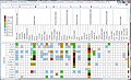 TumorGridWiki.jpg 1,890 × 1,148; 294 KB
-
 Tumour growing from the antrum Wellcome L0062372.jpg 3,907 × 5,372; 4.57 MB
-
 Tumours of uncertain nature in an upper arm Wellcome L0061237.jpg 4,124 × 5,616; 3.57 MB
-
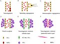 Types of Tumor Angiogenesis.jpg 778 × 577; 88 KB
-
 Blood vessel tumour, with perficial lesions in a female pati Wellcome V0036314EB.jpg 2,988 × 1,223; 1.22 MB
-
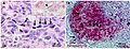 Vascular mimicry melanoma staining.jpg 483 × 184; 123 KB
-
 Vasculogenic mimicry signaling pathways.jpg 567 × 417; 64 KB
-
 Venous lake 1.jpg 512 × 336; 17 KB
-
 Venous lake 2.jpg 512 × 336; 19 KB
-
 Venous lake 3.jpg 512 × 336; 15 KB
-
 Vista frontale fondazione G. Pascale.jpg 500 × 375; 116 KB
-


_55-o.apatho-806a-tmoucuisse.jpg)
_55-o.apatho-806p-tmoucuisse.jpg)


