Category:Electrophysiology
Перайсці да навігацыі
Перайсці да пошуку
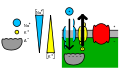
study of the electrical properties of biological cells and tissues. | |||||
| Запампаваць медыя | |||||
| Гэта | |||||
|---|---|---|---|---|---|
| Абагульняецца | |||||
| Частка ад | |||||
| |||||
Падкатэгорыі
Гэтая катэгорыя мае наступныя 33 падкатэгорый, з наяўных 33.
A
B
- BITalino (7 F)
- Brain electrophysiology (31 F)
C
E
- Electrocorticography (8 F)
- Electromyoneurography (1 F)
- Electrooculography (1 F)
- Electrophysiological phenomena (16 F)
- Electroreception (41 F)
- Evgeny Pokushalov (4 F)
I
L
- Radosław Lenarczyk (1 F)
M
- Microelectrodes (11 F)
- Muscle electrophysiology (15 F)
N
- Neural conduction (4 F)
- Neural oscillation (7 F)
- Neuron resting potential (6 F)
P
- Patch-clamp techniques (120 F)
S
- Spike sorting (1 F)
T
- Theta rhythm (4 F)
V
- Voltage clamps (15 F)
Мультымедыя ў катэгорыі "Electrophysiology"
У гэтай катэгорыі ёсць наступныя 167 файлаў з агульнага ліку 167.
-
 1P cultured neuron.png 582 × 686; 61 KB
-
 1P HEK293.png 618 × 671; 66 KB
-
 1P hESC-CM.png 711 × 518; 77 KB
-
 1P iPSC-CM.png 741 × 554; 77 KB
-
 2P sliceCulture 50usDwell.png 785 × 741; 105 KB
-
 3-D Mamma.jpg 725 × 595; 81 KB
-
 3274094 sensors-11-00310f6.png 484 × 195; 22 KB
-
 Acaide505f01th.jpg 450 × 448; 64 KB
-
 Additive response.png 532 × 423; 52 KB
-
 Aksiyon Potansiyeli.png 491 × 485; 11 KB
-
 Alci clinical picture.jpg 1 200 × 679; 124 KB
-
 Amp response.png 532 × 423; 58 KB
-
 Ampulla of Lorenzini electrophysiology.svg 720 × 983; 16 KB
-
 Analyse spectrale d'un EEG.jpg 1 156 × 1 772; 361 KB
-
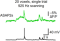 ASAP2s 2P sliceCulture single trial trace.png 754 × 513; 65 KB
-
 Der Tod durch Elektricität - eine forensisch-medicinische Studie auf experimenteller Grundlage (IA b21711070).pdf 1 122 × 1 687, 184 старонак; 10,32 MB
-
 Bastonetes e células bipolares no claro e no escuro eletrofisiologia.png 1 360 × 768; 91 KB
-
 Bioelectric potential modeled by Xenopus.png 1 024 × 768; 736 KB
-
 Bioelectricity Figure 1.png 853 × 602; 184 KB
-
 BMSEED’s electrophysiology module Intan 120-Channel.png 2 016 × 1 512; 2,52 MB
-
 BuenoOrovio2.png 1 855 × 1 001; 77 KB
-
 BuenoOrovioModel.png 1 855 × 1 001; 76 KB
-
 Bundleofhis.png 400 × 483; 69 KB
-
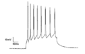
 Bursting-recording.png 661 × 197; 27 KB
-
 Calcium fluorescence imaging schematic.png 718 × 945; 82 KB
-
 Canais de sodio e potassio despolarizacao hiperpolarizacao.png 1 418 × 770; 140 KB
-
 Circuit electrique equivalent cellule.gif 960 × 720; 6 KB
-
 ColourDopplerA.jpg 400 × 273; 69 KB
-
 Corticography recording.png 444 × 355; 427 KB
-
 Corticotroph traces.png 2 481 × 1 057; 157 KB
-
 CTWristImage.png 798 × 636; 315 KB
-
 Current Clamp recording of Neuron.GIF 840 × 531; 8 KB
-
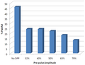
 Depolarizing Prepulse.PNG 896 × 601; 34 KB
-
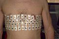
 Desfibrilador público.jpg 778 × 159; 130 KB
-

-
-
 Dup15q EEG margins.png 2 497 × 1 533; 934 KB
-
 Dup15q EEG signature.png 1 597 × 756; 499 KB
-
 EIT electrodes on chest Oxford Brookes.jpg 300 × 200; 55 KB
-
 EIT image of chest from Oxford Brookes OXBACT.png 522 × 369; 127 KB
-
 Electricity in the Service of Medicine. Wellcome M0017542.jpg 3 450 × 3 179; 2,27 MB
-
 Electro-physiology (1896-98) (20586945304).jpg 950 × 858; 200 KB
-
 Electro-physiology (1896-98) (21022824389).jpg 428 × 604; 83 KB
-
 Electro-physiology (1896-98) (21199671442).jpg 1 160 × 532; 132 KB
-
 Electrocytes.pdf 1 006 × 808; 249 KB
-
 Electrocytes.svg 1 200 × 962; 32 KB
-
 Electronic nose.jpg 1 150 × 620; 87 KB
-
 Electroretinographhjdeviass2.jpeg 2 884 × 1 409; 589 KB
-
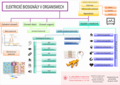 Elektrické biosignály v organismech.png 3 318 × 2 347; 1,35 MB
-
 Die Wissenschaft 044 Elektrobiologie. 1912 (IA elektrobiologied00bern).pdf 804 × 1 243, 278 старонак; 14,43 MB
-
 EOGhjdevias.jpeg 4 801 × 1 850; 357 KB
-
 Fibrosi postviral. Feix de His. 200X. Tricromic de Masson. IMG 2907.jpg 838 × 535; 120 KB
-
 Firing patterns.png 903 × 693; 442 KB
-
 Four images of experiment on the electro-physiological Wellcome L0035280.jpg 3 896 × 1 240; 816 KB
-
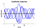
 Freq response.png 532 × 423; 84 KB
-
 Galvani, experiments in electrophysiology Wellcome L0002976.jpg 1 668 × 1 192; 1 016 KB
-
 Gibbs-donnan-de.svg 550 × 400; 175 KB
-
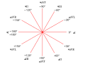 Hexaxial reference system.svg 718 × 539; 11 KB
-
 HFO wiki.jpg 1 534 × 684; 79 KB
-
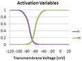 Hodgkin Huxley Model Activation Variables.PNG 470 × 352; 13 KB
-
 HRIC Atrium.JPG 2 048 × 1 536; 638 KB
-

-
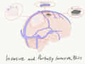 Invasive and partially invasive BCIs.png 2 048 × 1 536; 555 KB
-
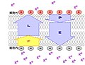
 Ion concentrations.svg 504 × 288; 135 KB
-
 Ion transport.svg 447 × 253; 66 KB
-
 Ionic equilibrium potential1.svg 550 × 400; 173 KB
-
 Ionic equilibrium potential2.svg 550 × 400; 222 KB
-
 Ionic equilibrium potential3.svg 550 × 400; 233 KB
-
 Ionrecept.jpg 556 × 290; 51 KB
-
 Josephson.png 226 × 442; 132 KB
-
 KEquilibrium.jpg 737 × 563; 176 KB
-
 Known age-related electrophysiological and morphological changes in neurons.jpg 950 × 1 454; 579 KB
-
 LCIA Sign.jpg 2 048 × 1 536; 711 KB
-
 Les récepteurs membranaires comme cibles thérapeutiques.png 3 184 × 2 050; 163 KB
-
 Levin Figure 5.png 482 × 613; 187 KB
-
 Levin Figure 7.png 1 024 × 768; 251 KB
-
 Limiar e intensidade do sinal sensorial.png 1 528 × 543; 49 KB
-
 LPP for emotional relevant VS irrelevant facial expression.jpg 850 × 754; 180 KB
-
 Mamma ca 1.jpg 497 × 344; 55 KB
-
 Mamma ca farbe.jpg 505 × 341; 65 KB
-
 Manually Stretching Microelectrode Array.jpg 1 920 × 1 080; 805 KB
-
 Mauthner Cell axon cap schematic.svg 325 × 295; 23 KB
-
 Membrane electric profile.svg 512 × 422; 1 KB
-
 Membrane Ions.png 1 075 × 563; 158 KB
-
 Membrane Permeability of a Neuron During an Action Potential.pdf 1 500 × 1 125; 66 KB
-
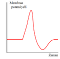
 Membrane potential development.jpg 1 619 × 518; 79 KB
-
 Membrane potential ions (id).jpg 550 × 400; 186 KB
-
 Membrane potential ions en.svg 550 × 400; 185 KB
-
 Membrane potential ions it.svg 550 × 400; 185 KB
-
 Membrane potential.jpg 1 024 × 589; 278 KB
-
 Metallic arc used by Luigi Galvani, Europe, 1775-1798 Wellcome L0057741.jpg 4 050 × 2 226; 732 KB
-
 MFI-system-wiki.jpg 400 × 620; 53 KB
-
 Microscope for Electrophysiological Research and Recording Equipment.jpg 2 288 × 1 712; 645 KB
-
 Microscope for Electrophysiological Research shielded by Faraday Cage - (1).jpg 1 712 × 2 288; 556 KB
-
 Microscope for Electrophysiological Research shielded by Faraday Cage - (2).jpg 1 712 × 2 288; 656 KB
-

-
 Microscope for Electrophysiological Research shielded by Faraday Cage.jpg 2 288 × 1 712; 693 KB
-
 MinZhaoImage1.jpg 878 × 271; 33 KB
-
 MinZhaoImage2.jpg 593 × 520; 68 KB
-
 MRS spectrum.gif 395 × 282; 6 KB
-
 Muskulatur - Einzelzuckung ar.PNG 1 123 × 805; 40 KB
-
 Muskulatur - Einzelzuckung.png 1 123 × 805; 38 KB
-
 Muskulatur - unvollstaendiger Tetanus ar.PNG 1 123 × 804; 44 KB
-
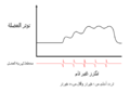
 Muskulatur - unvollstaendiger Tetanus.png 1 123 × 804; 45 KB
-
 Muskulatur - vollstaendiger Tetanus ar.PNG 1 123 × 805; 54 KB
-
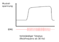 Muskulatur - vollstaendiger Tetanus.png 1 123 × 805; 50 KB
-
 NCS f-wave.gif 2 080 × 548; 252 KB
-
 NCS peronaeus.gif 2 080 × 548; 247 KB
-
 NCS suralis.gif 2 080 × 548; 238 KB
-
 Nothing Downstream.png 697 × 530; 219 KB
-
 PET-IRM-cabeza-Keosys.JPG 1 280 × 1 024; 117 KB
-
 Phase resetting.png 532 × 423; 69 KB
-
 Phase space trajectory of FitzHugh-Nagumo model.svg 489 × 325; 52 KB
-
 Pipette Puller-de.jpg 4 209 × 3 175; 262 KB
-
 Pipette Puller-de.svg 600 × 600; 40 KB
-
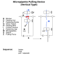 Pipette Puller-en.svg 600 × 600; 46 KB
-
-
 Poletto 2002 pain plot 2.PNG 777 × 595; 21 KB
-
 Portable heart rate variability device.JPG 1 667 × 2 500; 841 KB
-
 Potassium equilibrium.svg 757 × 781; 124 KB
-
 Potential registration.svg 1 026 × 507; 39 KB
-
 Resting potential.svg 160 × 100; 25 KB
-
 Rheobase chronaxie.png 872 × 510; 32 KB
-
 Rheobase chronaxie.svg 734 × 363; 8 KB
-
 RTI-32 structure.png 802 × 482; 10 KB
-
 Salle de vasculaire.jpg 1 280 × 1 024; 168 KB
-
 Science edunihgovinfo fig02.gif 500 × 292; 11 KB
-
 Selektiivne isolatsioon.svg 1 052 × 744; 15 KB
-
 Shapes of the cardiac action potential in the heart.svg 1 000 × 750; 201 KB
-
 Simulation of variable x(t) in Fitzhugh-Nagumo model.svg 485 × 323; 22 KB
-
 Simulation of variable x(t) in Hindmarh-Rose model.svg 492 × 329; 27 KB
-
 Sodium equilibrium.svg 249 × 262; 48 KB
-
 Spike clusters.png 397 × 319; 2 KB
-
 Spike cutouts sorted.png 444 × 347; 13 KB
-
 Spike triggered averages.png 800 × 830; 86 KB
-
 Stretchable Microelectrode Array (sMEA) before and during stretch.png 2 205 × 1 404; 3,42 MB
-
 Stretchable Microelectrode Array (sMEA) with PDMS glue.jpg 3 024 × 4 032; 1,7 MB
-
 Stretchable Microelectrode Array (sMEA) with white glue.jpg 2 732 × 2 732; 1,31 MB
-
 Supplemental Fig. 2.png 2 550 × 2 550; 2,69 MB
-
 Surround suppression revised.png 966 × 691; 93 KB
-
 SVT2012.JPG 4 248 × 2 144; 3 MB
-
-
-
-
-
 Tomograf matryca voxele.png 551 × 389; 12 KB
-
 Tomograf metoda sumacyjna z filtr.png 523 × 512; 18 KB
-
 Topologie van het humane Kir2.2-ionkanaaleiwit.png 1 706 × 2 439; 360 KB
-
 UCA generation 1.jpg 143 × 143; 5 KB
-
 UltrasoundProbe2006a.jpg 400 × 247; 36 KB
-

-
 Ussingchamber.png 1 415 × 1 192; 41 KB
-
-
-
-
-
-
-
-
-
-
-
 Vascular examination.png 881 × 1 007; 950 KB
-
 Volume conductor model.png 466 × 327; 261 KB
-
 Weakly Electric Fish Navigating Electric Fields.jpg 434 × 189; 24 KB
-
 Микроэлектрод.png 1 622 × 2 482; 4,67 MB
-
 창원파티마병원 혈관조영실.jpg 4 256 × 2 832; 4,4 MB


_(14597337730).jpg)


_(20586945304).jpg)
_(21022824389).jpg)
_(21199671442).jpg)


_to_allow_electrophysiological_output_recording.jpg)


.jpg)


.jpg)
.jpg)


_(14764260585).jpg)


_in_Fitzhugh-Nagumo_model.svg)
_in_Hindmarh-Rose_model.svg)


_before_and_during_stretch.png)
_with_PDMS_glue.jpg)
_with_white_glue.jpg)


