Category:Ion channels
Jump to navigation
Jump to search
pore-forming membrane protein that allows the passage of ions through a membrane  | |||||
| Upload media | |||||
| Instance of |
| ||||
|---|---|---|---|---|---|
| Subclass of |
| ||||
| |||||
Subcategories
This category has the following 17 subcategories, out of 17 total.
Media in category "Ion channels"
The following 52 files are in this category, out of 52 total.
-
 2006 - Rob Flewell.jpg 921 × 1,024; 251 KB
-
 Alamethicin ion channel.jpg 250 × 250; 18 KB
-
 Blausen 0225 ChemicallyGatedChannel.png 1,500 × 2,500; 3.36 MB
-
 Cell IV showing rest and AP thresh.png 600 × 444; 7 KB
-
 Currents responsible for the cardiac action potential.png 960 × 720; 62 KB
-
 Difference between voltage-gated and ligand-gated channels.png 344 × 599; 38 KB
-
 F42 K+channel select stereo.jpg 2,000 × 1,019; 597 KB
-
 ΔFosB.svg 935 × 965; 238 KB
-
 Gramicidin A.gif 256 × 256; 547 KB
-
 Heteroscodratoxin-1.jpg 500 × 500; 24 KB
-
 HilleEqnParameters.svg 244 × 311; 301 KB
-
 Ion channel activity before during and after polarization.jpg 1,117 × 1,583; 735 KB
-
 Ion channel image - Kim 2014 PMCID 3935107.png 1,290 × 688; 535 KB
-
 Ion channel in conformation.png 378 × 450; 28 KB
-
 Ion channel.png 705 × 661; 99 KB
-
 Ion Channel.png 1,064 × 552; 215 KB
-
 Ion channels.png 863 × 268; 74 KB
-
 Ion channels.svg 374 × 651; 41 KB
-
 Ivermectin mechanism of action 3RHW.png 3,250 × 2,900; 2.57 MB
-
 Kanalprotein.png 796 × 497; 26 KB
-
 Mechanoelectrical transduction in hair cells.png 3,858 × 2,699; 1.28 MB
-
 Misexpression of ion channels.crop.png 345 × 409; 111 KB
-
 Misexpression of ion channels.png 1,024 × 768; 138 KB
-
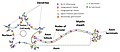
 Na-Glucose-Symport.svg 942 × 535; 158 KB
-
 Neuron with ALL channels.jpg 1,498 × 654; 137 KB
-
 Neutron diffraction; Ion channels (5888008521).jpg 1,016 × 662; 551 KB
-
 NMDA Receptor.png 2,808 × 1,572; 94 KB
-
 Open and closed conformations of ion channels.png 1,038 × 746; 268 KB
-
 PBB Protein ITPR1 image.jpg 500 × 500; 40 KB
-
 PBB Protein KCNH2 image.jpg 500 × 500; 30 KB
-
 PBB Protein RYR1 image.jpg 434 × 381; 43 KB
-
 Prokaryotic stretch-activated channel.jpg 1,008 × 630; 79 KB
-
 Protein gates.png 640 × 480; 9 KB
-
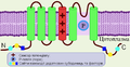
 Récepteur ionotrope.svg 606 × 620; 339 KB
-
 Schematic illustration Piezo1-channel, closed-open conformation..jpg 1,026 × 1,376; 165 KB
-
 SCNN1G-gene-transcript-Hanukoglu.png 956 × 202; 21 KB
-
 SK Channel.jpg 800 × 600; 124 KB
-
Slo1-is-the-principal-potassium-channel-of-human-spermatozoa-elife01009v001.ogv 11 s, 288 × 384; 242 KB
-
Slo1-is-the-principal-potassium-channel-of-human-spermatozoa-elife01009v002.ogv 11 s, 288 × 384; 255 KB
-
 Sodium calcium pump.png 1,422 × 951; 30 KB
-
 Sodium Channels Open Closed Depolarization.jpg 321 × 599; 44 KB
-

-
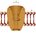
 Structure of a Prokaryotic CNG Channel.jpg 1,270 × 1,398; 876 KB
-
 Structure of a Prokaryotic CNG Channel.pdf 1,322 × 1,456; 15.14 MB
-
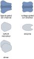
 Subunits of ion channels in membrane.png 1,040 × 1,032; 177 KB
-
Synapsis.theora.ogv 1 min 0 s, 640 × 480; 12.2 MB
-
 Thermoreception 1.png 4,236 × 2,812; 1.12 MB
-
 Thermoreception 2.png 4,236 × 2,812; 1.18 MB
-
 Vesicle technique.svg 876 × 683; 1.17 MB
-
 Voltage-clamp-concept.svg 1,078 × 1,368; 9.58 MB
-
 ΔFosB vi.svg 935 × 965; 245 KB
-
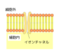 イオンチャネルの模式図.svg 800 × 750; 95 KB


.jpg)


_and_soluble-form_PVL_(pale_green-green)_toxins_-_PDB_7AHL_and_1T5R.png)


