Category:Human spinal cord
Vai alla navigazione
Vai alla ricerca
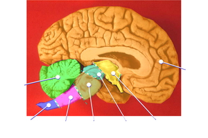
Human brain: sagittal section
|
spazio vertebrale  | |||||
| Carica un file multimediale | |||||
| Istanza di |
| ||||
|---|---|---|---|---|---|
| Sottoclasse di |
| ||||
| Parte di | |||||
| |||||
Sottocategorie
Questa categoria contiene le 11 sottocategorie indicate di seguito, su un totale di 11.
File nella categoria "Human spinal cord"
Questa categoria contiene 98 file, indicati di seguito, su un totale di 98.
-
 1982 Decompression sickness 1.JPG 500 × 504; 97 KB
-
 1982 Decompression sickness 2.JPG 777 × 509; 147 KB
-
 78 MAV Peri-medullaire1.jpg 1 876 × 2 530; 681 KB
-
-

-
 Artery of Adamkiewicz CT scan OsiriX.jpg 640 × 720; 81 KB
-
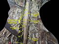
 Basel 2012-10-05 Batch 2 (30).JPG 2 736 × 3 648; 2,86 MB
-
 Blausen 0822 SpinalCord.png 768 × 1 024; 2,25 MB
-
 Bourgery & Jacob-r20.jpg 2 847 × 3 969; 3,16 MB
-

-
 Brain and spinal cord; dissection, back view. Coloured line Wellcome V0008396.jpg 2 055 × 3 406; 3,53 MB
-
 Braus 1921 52.png 1 640 × 2 556; 12,01 MB
-
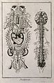
 C. J. M. Langenbeck, Icones anatomicae. Wellcome L0022123.jpg 1 232 × 1 550; 459 KB
-
 C. J. M. Langenbeck, Icones anatomicae; Wellcome L0022125.jpg 1 224 × 1 574; 318 KB
-
 Cerebro-spinal Fluid cavity at bottom of spine.JPG 250 × 500; 27 KB
-
 Ch13 vertebral column.png 543 × 695; 209 KB
-
-

-
 Diagram of the Spinal Cord Unlabeled.jpg 1 698 × 2 523; 412 KB
-
 Differentiating intramedullary pathology, location within the cord.webp 1 524 × 816; 145 KB
-
 Dissection of spinal cord.jpg 960 × 720; 87 KB
-
 Embryonic spinal cord.jpg 457 × 277; 21 KB
-
 Flatau prawo 1897.jpg 1 342 × 1 900; 442 KB
-
 Gray1213.png 250 × 500; 18 KB
-
 Gray661.png 311 × 550; 33 KB
-
 Gray663-hu.jpg 396 × 1 125; 81 KB
-
 Gray663.png 264 × 750; 13 KB
-
 Gray796.png 291 × 500; 39 KB
-
 Gray839 nippon.png 479 × 700; 71 KB
-
 Gray839-d.png 893 × 1 260; 828 KB
-
 Gray839-zh.png 479 × 700; 154 KB
-
 Gray839.png 479 × 700; 61 KB
-
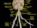
 Human brain and spinal cord.jpg 1 003 × 3 232; 1,33 MB
-
 Human spinal cord anterior view.jpg 661 × 461; 28 KB
-
 J. M. Charcot, Diseases of the nervous syste Wellcome L0029910.jpg 2 541 × 4 332; 4,26 MB
-
 J. M. Charcot, Diseases of the nervous syste Wellcome L0029911.jpg 2 590 × 4 224; 4,57 MB
-
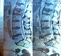
 Lesionmedula.jpg 399 × 438; 51 KB
-
 Liblikas, mis ei oska lennata.jpg 1 187 × 698; 318 KB
-
 Lower spinal cord (expanded).gif 60 × 409; 9 KB
-
 Lower spinal cord.gif 30 × 150; 4 KB
-
 MAVmedul10.jpg 1 200 × 1 600; 306 KB
-
 Meyers b12 s0057a.jpg 2 048 × 1 675; 670 KB
-
 Meyers b12 s0057b.jpg 2 048 × 1 657; 636 KB
-
 Moelle Epinière.png 482 × 370; 55 KB
-
 Monakow atlas.JPG 614 × 1 582; 192 KB
-
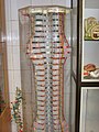 Moulage of spinal cord segments.JPG 2 112 × 2 816; 2,87 MB
-
 Nervosraq.jpg 165 × 252; 10 KB
-
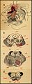
 Nervous system after Vieussens (fig. 1); brain and spinal co Wellcome V0007827.jpg 2 125 × 3 324; 3,26 MB
-
 Nervous system after Vieussens (fig. 1); brain and spinal co Wellcome V0007841.jpg 2 244 × 3 449; 3,36 MB
-
 Nervous system; five figures, showing the nerves, brain and Wellcome V0008027ER.jpg 1 181 × 1 574; 1,21 MB
-
 Nervous system; five figures, showing the nerves, brain and Wellcome V0008033EL.jpg 1 169 × 1 557; 1,23 MB
-
 New Mixed Myeloid-Lymphoid Progenitor Tree(RCCH) Grayscale.jpg 1 268 × 587; 178 KB
-
 Nucleus of onuf.PNG 1 274 × 1 658; 104 KB
-
 Onurğa beyni at quyruğu.jpg 400 × 320; 82 KB
-

-
 P1120690a-001.JPG 2 608 × 2 412; 2,61 MB
-
 P1120691a.JPG 2 303 × 2 144; 1,74 MB
-
 Posterior view of human spinal cord (1).jpg 2 288 × 1 520; 1,28 MB
-
 Rückenmark - 4738.jpg 2 692 × 12 392; 29,39 MB
-
SAG.-T2-FRFSE-L-SPINE.ogg 5,3 s, 1 362 × 896; 468 KB
-
 Several examples of diseased brain and spina Wellcome V0009856.jpg 648 × 486; 81 KB
-
 Siebert 03.jpg 1 962 × 2 866; 1,62 MB
-
 Slide12ee.JPG 960 × 720; 138 KB
-
 Slide1drdr.GIF 960 × 720; 459 KB
-
 Slide2drdr.GIF 960 × 720; 452 KB
-
 Slide2fer.JPG 960 × 720; 93 KB
-
 Slide2MIR.JPG 960 × 720; 115 KB
-
 Slide2PIT.JPG 960 × 720; 97 KB
-
 Slide2PITER.JPG 960 × 720; 77 KB
-
 Slide2ZEO-ar.jpg 960 × 720; 169 KB
-
 Slide2ZEO.JPG 960 × 720; 82 KB
-
 Slide3dsdd.GIF 960 × 720; 367 KB
-
 Slide3fer.JPG 960 × 720; 79 KB
-
 Slide3PIT.JPG 960 × 720; 105 KB
-
 Slide3ZEO.JPG 960 × 720; 93 KB
-
 Slide4rer.JPG 960 × 720; 77 KB
-
 Slide5rer.JPG 960 × 720; 102 KB
-
 Spinal Compression Pain.png 768 × 1 024; 692 KB
-
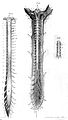
 Spinal Cord - 4742.jpg 785 × 4 139; 2,95 MB
-
 Spinal cord and roots and dural tube which covers them. Wellcome L0002010.jpg 1 026 × 1 810; 518 KB
-
 SPINAL CORD BLEED.GIF 159 × 160; 10 KB
-
 Spinal cord stimulator.jpg 792 × 1 006; 411 KB
-
 Spinal cord.gif 99 × 1 013; 21 KB
-
 Spinal cord.jpg 640 × 480; 37 KB
-
 Spinal Graft Displacement.png 764 × 1 024; 725 KB
-
 Spinal readjustment 1.jpg 411 × 481; 24 KB
-
 Spinal readjustment 2.jpg 492 × 562; 41 KB
-
 Spinal Stenosis.png 768 × 1 024; 2,25 MB
-
 Spinalnerve.png 300 × 236; 12 KB
-
 Spine Mechanical Pain.png 764 × 1 024; 748 KB
-
 Spine, ribcage and pelvis, with eight figures illustrating v Wellcome V0008175EL.jpg 1 104 × 1 716; 1,09 MB
-
 Spinewithcord.jpg 275 × 212; 45 KB
-
 The human brain and associated nerves Wellcome L0074578.jpg 4 169 × 5 922; 6,61 MB
-
 The spinal medula, cut transversly. Wellcome L0001573.jpg 1 388 × 1 394; 939 KB
-
 Traité des maladies de la moelle épinière (1902) (14595390079).jpg 1 576 × 2 474; 557 KB
-

-
 Ön və yan piramid yollar.gif 566 × 706; 55 KB
-
 طناب نخاعی.jpeg 114 × 216; 9 KB


_(14769058655).jpg)


.JPG)


_(14577756788).jpg)


;_brain_and_spinal_co_Wellcome_V0007827.jpg)
;_brain_and_spinal_co_Wellcome_V0007841.jpg)


_Grayscale.jpg)


_(14758612216).jpg)


.jpg)


_(14595390079).jpg)
_(14784768063).jpg)


.gif)