Category:Human brain
Jump to navigation
Jump to search
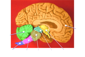
Human brain: sagittal section
|
Français : Cerveau humain
Kurdî: Mejiyê mirov
main organ of the human nervous system   | |||||
| Upload media | |||||
| Spoken text audio | |||||
|---|---|---|---|---|---|
| Instance of |
| ||||
| Subclass of |
| ||||
| Part of | |||||
| Has part(s) | |||||
| Mass |
| ||||
| Follows | |||||
| Different from | |||||
| |||||
Subcategories
This category has the following 31 subcategories, out of 31 total.
A
- Albert Einstein's brain (1 F)
B
D
E
- Brain electrophysiology (31 F)
L
M
- Memoirs (1783) (5 F)
N
P
S
- SpiNNaker (3 F)
T
V
- Brain in a vat (1 P, 44 F)
Y
- Yakovlevian torque (1 F)
Media in category "Human brain"
The following 150 files are in this category, out of 150 total.
-
 15 intelligence knowing-neurons.jpg 838 × 1,024; 61 KB
-
 2013 seli-seli-seppele-yleiskuva ore-e-refineries c-none q xxx e-1-a z img.JPG 4,416 × 3,312; 8.25 MB
-
7 Tesla MRI of the ex vivo human brain at 100 micron resolution (axial, coronal, sagittal and descriptive summary).webm 4 min 56 s, 1,920 × 1,080; 69.97 MB
-
 9727513.fig.jpg 600 × 255; 29 KB
-
 A brain, viewed from the top, supposedly from a subject with Wellcome V0030015.jpg 2,279 × 3,090; 1.65 MB
-
 A cerebral atlas Wellcome L0070279.jpg 5,701 × 6,980; 7.73 MB
-
 A cerebral atlas Wellcome L0070280.jpg 5,654 × 6,980; 7.72 MB
-
 Acetylcholine Pathway-es.png 1,400 × 991; 275 KB
-
 Acetylcholine Pathway.png 1,350 × 955; 1.01 MB
-

-
 Afni oblique.png 424 × 450; 43 KB
-
 Allen brain atlas.png 532 × 172; 62 KB
-
 Amygdala dorsal view.png 767 × 932; 430 KB
-
 An example of unparalleled illustrations of the Atlas .png 806 × 772; 849 KB
-
 Ape NE cells undergo cell shape transition before the onset of neurogenesis.jpg 3,333 × 2,876; 1.44 MB
-
 Aprenentatge.jpg 3,555 × 2,554; 374 KB
-
 Aprosodieross.jpg 734 × 454; 145 KB
-
 Ars Longa by Sholim.gif 167 × 250; 688 KB
-
 Attempt to use human brain to receive radio waves.jpg 629 × 457; 91 KB
-
 Autismbrain TR.jpg 400 × 602; 176 KB
-
 Brain AS.stl 5,120 × 2,880; 145.77 MB
-
 Brain Exercising.png 1,920 × 1,440; 532 KB
-
 Brain Lateralization.png 1,920 × 1,024; 973 KB
-
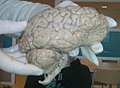
 Brain model 12.jpg 3,872 × 2,592; 3.33 MB
-
 Brain of marijuana user.jpg 400 × 302; 9 KB
-
 Brain Racism Complex.jpg 617 × 482; 104 KB
-
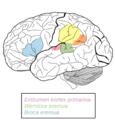
 Brain regions involved in memory formation.jpg 532 × 306; 30 KB
-
 Brain structure biology.jpg 2,744 × 3,659; 1.36 MB
-
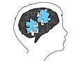 Brain with a head outline and grey and blue puzzle pieces.jpg 720 × 540; 40 KB
-
 Brain-954822 1280.jpg 3,849 × 2,974; 2.36 MB
-
 Brain-Pull-CIS-2012-12.png 890 × 986; 72 KB
-
 Brainstem AS.stl 5,120 × 2,880; 16.84 MB
-
 Cerebellum AS.stl 5,120 × 2,880; 22.27 MB
-
 CIT168 T1w 700um4.png 514 × 514; 134 KB
-
 Decisionbrain.jpg 373 × 221; 79 KB
-
 Delayed human NE transition is associated with a shorter cell cycle.jpg 2,922 × 4,169; 1.4 MB
-
 Diencephalic Syndrome 5.jpg 814 × 811; 258 KB
-
 Diencephalic Syndrome 6.jpg 825 × 815; 252 KB
-
 DrPaulineNeveu 02 Systemes 5HT.svg 975 × 653; 892 KB
-
 DrPaulineNeveu 02 Systemes A.svg 975 × 653; 875 KB
-
 DrPaulineNeveu 02 Systemes Ach.svg 975 × 653; 887 KB
-
 DrPaulineNeveu 02 Systemes DA.svg 976 × 655; 886 KB
-
 DrPaulineNeveu 02 Systemes HA.svg 975 × 653; 883 KB
-
 DrPaulineNeveu 02 Systemes NA.svg 975 × 653; 888 KB
-
 Dup15q EEG signature.png 1,597 × 756; 499 KB
-
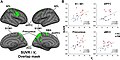 Fibromyalgia Glial Activation Agreement between SUVR and VT analyses A. Fig. 3.jpg 2,159 × 1,076; 237 KB
-

-
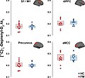
 Fibromyalgia Glial Activation Voxelwise group differences in (11C)PBR28 VT. A Fig. 1.jpg 2,130 × 1,104; 237 KB
-
-

-
 Figure01 BEM sources revised.png 3,191 × 2,615; 1.24 MB
-
 Figure03 Unconstrained Resolution rev.png 1,675 × 2,506; 1.44 MB
-
 Figure03 Unconstrained Resolution.png 1,675 × 2,506; 1.44 MB
-
 Flaticonbrain.png 733 × 711; 28 KB
-
 Free 3D Illustration Of A Mental Health Conceptual Image By Quince Media 01.jpg 5,000 × 2,750; 1.41 MB
-
 Free 3D Illustration Of A Mental Health Conceptual Image By Quince Media 02.jpg 5,000 × 2,750; 1.26 MB
-
 Free 3D Illustration Of A Mental Health Conceptual Image By Quince Media 03.jpg 5,000 × 2,750; 1.36 MB
-
 Free 3D Illustration Of A Mental Health Conceptual Image By Quince Media 04.jpg 5,000 × 2,750; 1.32 MB
-
 Free 3D Illustration Of A Mental Health Conceptual Image By Quince Media 05.jpg 5,000 × 2,750; 2.22 MB
-
 Free 3D Illustration Of A Mental Health Conceptual Image By Quince Media 06.jpg 5,000 × 2,750; 1.2 MB
-
 Free 3D Illustration Of A Mental Health Conceptual Image By Quince Media 07.jpg 5,000 × 2,750; 1.34 MB
-
 Free 3D Illustration Of A Mental Health Conceptual Image By Quince Media 08.jpg 5,000 × 2,750; 1.81 MB
-
 Free 3D Illustration Of A Mental Health Conceptual Image By Quince Media 09.jpg 5,000 × 2,750; 814 KB
-
 Free 3D Illustration Of A Mental Health Conceptual Image By Quince Media 10.jpg 5,000 × 2,750; 1.84 MB
-
 Free 3D Illustration Of A Mental Health Conceptual Image By Quince Media 11.jpg 5,000 × 2,750; 1.38 MB
-
 Free 3D Illustration Of A Mental Health Conceptual Image By Quince Media 12.jpg 5,000 × 2,750; 2.29 MB
-
 Free 3D Illustration Of A Mental Health Conceptual Image By Quince Media 13.jpg 5,000 × 2,750; 3.46 MB
-
 Frequency mapping in human ear and brain - 10.1371 journal.pbio.0030137.g001-L.jpg 2,020 × 2,480; 989 KB
-
 Friern Hospital, London; a brain, seen from beneath. Photogr Wellcome V0029602.jpg 2,560 × 2,932; 2.7 MB
-
 Friern Hospital, London; a brain, seen from underneath. Phot Wellcome V0029604.jpg 3,040 × 2,552; 3.76 MB
-
 Friern Hospital, London; a brain. Photograph, 1890-1910. Wellcome V0029605.jpg 3,124 × 2,444; 2.94 MB
-
 Friern Hospital, London; a diseased brain, viewed from above Wellcome V0029645.jpg 2,888 × 2,516; 2.74 MB
-

-
 From brain to wikipedia.png 1,000 × 1,000; 402 KB
-
 Füüsilise antropoloogia pildistus.jpg 9,952 × 7,273; 29.68 MB
-
 Galvos smegenys (šizofrenija). Iliustracijos..png 960 × 720; 285 KB
-
 Galvos smegenys (šizofrenija). Iliustracijos.1.png 957 × 617; 313 KB
-
 Genetic correlations of brain imaging traits and language skill-levels.jpg 2,024 × 662; 182 KB
-

-
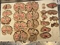 Gross pathology of brain after sectioning.jpg 2,869 × 2,117; 1.68 MB
-
 Haley Scans 071205 19892.jpg 450 × 300; 25 KB
-
 Head divided into four cerebral lobes; profile. Drawing with Wellcome V0009518.jpg 2,542 × 3,422; 3.36 MB
-
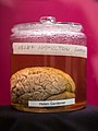 Helen Hamilton Gardener - Wilder Brain Collection.jpg 2,257 × 3,000; 2.58 MB
-
 Helen Hamilton Gardener and her brain Wellcome L0070273.jpg 7,666 × 5,253; 11.65 MB
-
 Help-Seeking is Cool.jpg 2,560 × 1,587; 830 KB
-
 Hjerne med brainstem.jpg 1,110 × 675; 99 KB
-
 Hjerne.png 1,362 × 898; 595 KB
-
 Hjärnan.jpg 275 × 183; 10 KB
-
 HoldBrainStitch.jpeg 2,433 × 2,134; 1.6 MB
-
 How music affects the brain (HY).svg 512 × 288; 40 KB
-
-
Human Brain Circumventricular Organs 6 - Subcommissural Organ - Sanjoy Sanyal.webm 1 min 10 s, 352 × 200; 4.57 MB
-
 Human brain in a vat.jpg 1,706 × 2,155; 1.3 MB
-
 Human NE cells exhibit species-specific differences in cell shape.jpg 3,329 × 2,942; 1.58 MB
-
 Human telencephalic organoids are larger with extended apical lumens.jpg 2,875 × 3,556; 1.17 MB
-
 Humanbraaiin.jpg 855 × 927; 383 KB
-
 Illustration of the brain Wellcome L0050374.jpg 3,723 × 4,728; 3.77 MB
-
 Illustrations of processes of mummification Wellcome M0008471.jpg 3,200 × 3,492; 2.15 MB
-

-
 Journal.pone.0052528.g001.png 1,661 × 1,160; 883 KB
-
 Adult male brain - natural size Wellcome L0001030EB.jpg 1,455 × 1,318; 678 KB
-
 Left right brain.jpg 1,667 × 1,667; 615 KB
-
 Lily Allen gig Nottingham 2009 MMB 25 Professor Green.jpg 2,790 × 1,838; 1.92 MB
-
 Logo ccn white1.png 1,181 × 1,181; 61 KB
-
 Loki lobuluko eremuak.png 379 × 397; 68 KB
-
 ManiaALE.png 522 × 522; 100 KB
-
 Mapa d'expressió.png 2,000 × 2,000; 1.69 MB
-
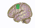
 Medialtemporallobe.jpg 2,272 × 1,704; 216 KB
-
 MIchelFerrari2009.jpg 1,733 × 2,600; 393 KB
-
 Navigation-search-brain.jpg 2,916 × 2,057; 996 KB
-
 Neurocranio.jpg 960 × 720; 64 KB
-
 Neurologische Reaktion bei Trauma, Stress und Tremor Response.pdf 1,239 × 1,754; 69 KB
-
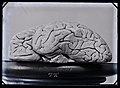
 NHM - Brain 2.jpg 800 × 600; 53 KB
-
 Oie vyqroZrtOsNl.png 900 × 900; 505 KB
-
 Partie du cerveau.gif 397 × 320; 17 KB
-
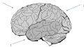
 Parts of the human brain.png 1,558 × 1,521; 390 KB
-
 Parts of the human cell.png 1,987 × 1,343; 303 KB
-
 Pavillon thématique L'homme et la vie, Expo 67.jpg 270 × 240; 32 KB
-
 Phineas Gage CGI.jpg 239 × 347; 25 KB
-
 Poke-a-brain.jpg 800 × 600; 124 KB
-
-
 Post-mortem pathology Wellcome L0070275.jpg 5,303 × 5,786; 14.01 MB
-
 PSM V35 D765 Diagram illustrating the concept rose.jpg 1,013 × 1,302; 85 KB
-
 PTSD.png 255 × 197; 62 KB
-
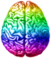 Rainbow brain, Aug 2014.png 420 × 480; 338 KB
-
 Rechterhelft Brein.jpg 1,023 × 752; 90 KB
-
 Reduced blood flow in cannabis users.jpg 432 × 352; 30 KB
-

-
 RR5110-0078R.gif 250 × 250; 48 KB
-
 Rysunek mózg zad23.svg 478 × 268; 311 KB
-
 Sir H. Head, Model of the brain, 1926 Wellcome L0015601.jpg 1,472 × 1,318; 785 KB
-
 Smegenų dalys, reguliuojančios kūno judesius.gif 543 × 767; 135 KB
-
 Smegenų dalys, siejamos su savikontrole ir judėjimu.gif 772 × 664; 163 KB
-
 Smegenų kraujotaka.jpg 677 × 431; 77 KB
-

-
 SN.tif 1,165 × 629; 1.53 MB
-
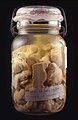
 Specimen jar containing piece of William Burke's brain Wellcome L0065696.jpg 2,832 × 4,256; 1.65 MB
-
 Spherical Registration.png 1,085 × 904; 664 KB
-
 Standard sections of brain.jpg 3,190 × 2,433; 1.77 MB
-
 Supplementary motor cortex.gif 400 × 312; 5.89 MB
-
 Terracotta votive scalp, Roman, 200 BCE-200 CE Wellcome L0057477.jpg 4,256 × 2,832; 1.84 MB
-
 The Journal of nervous and mental disease (1874) (14760534826).jpg 1,952 × 1,566; 237 KB
-
 The right hemisphere of the brain , cerebellum and brainstem AS.stl 5,120 × 2,880; 92.44 MB
-
 The right hemisphere of the brain AS.stl 5,120 × 2,880; 53.33 MB
-
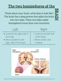 The two hemispheres.pdf 2,700 × 3,600, 2 pages; 208 KB
-
 The-spinal-cord-s-mcintosh.pdf 1,087 × 3,437; 1.25 MB
-
 Tomas Diaz MRI self portrait 01.jpg 1,380 × 1,784; 1.49 MB
-
 Vuyvem frontal 1.png 700 × 570; 139 KB
-
 Wax anatomical model of human head, Europe Wellcome L0059770.jpg 2,832 × 4,256; 1.29 MB
-


PBR28_SUVR_A._Fig._2.jpg)
PBR28_VT._A_Fig._1.jpg)


._Iliustracijos..png)
._Iliustracijos.1.png)


.svg)


.svg)


_(14760534826).jpg)


_v_Praze_(ilustracni_fotografie)_(UK0245).jpg)