Category:Immune system
Перайсьці да навігацыі
Перайсьці да пошуку
biological system | |||||
| Загрузіць мэдыя | |||||
| Асобны выпадак панятку |
| ||||
|---|---|---|---|---|---|
| Падкляса ад | |||||
| Складаецца з |
| ||||
| |||||
Падкатэгорыі
Гэтая катэгорыя зьмяшчае наступныя 38 падкатэгорыяў з 38 агулам.
A
- Acquired immune system (11 F)
B
C
E
G
- Graft survival (6 F)
H
- Haptens (3 F)
I
- Immune response (371 F)
- Immune tolerance (23 F)
- Immunosenescence (8 F)
- Immunotoxins (3 F)
- Innate immune system (53 F)
L
M
- MHC restriction (4 F)
P
S
T
- Transfer factor (4 F)
V
- Vernix caseosa (8 F)
Файлы ў катэгорыі «Immune system»
Паказаныя 143 файлы гэтай катэгорыі з 143.
-
 1aly.png 878 × 600; 124 кб
-
 1lkkA SH2 domain.png 948 × 1014; 233 кб
-
 2BC4.pdb1.png 1436 × 759; 274 кб
-
 2BVE.pdb.jpg 492 × 489; 41 кб
-
 2KLL.pdb.png 1436 × 759; 249 кб
-
 3hla ribbon.png 1000 × 1300; 98 кб
-
 3LTQ.pdb.png 1436 × 759; 173 кб
-
 Activation des lymphocytes T par un antigène conventionnel.jpg 1832 × 548; 94 кб
-
 Activation of B cells to make antibody.jpg 570 × 429; 30 кб
-
 Activation polyclonale des lymphocytes T par un superantigène.jpg 699 × 414; 44 кб
-
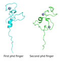 Aire protein (first- and second phd fingers).png 598 × 613; 71 кб
-
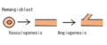 Angiogenesis.png 399 × 169; 8 кб
-
 Antibody and its corresponding antigen.svg 254 × 155; 33 кб
-
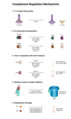
 Antibody Effector Mechanisms.png 5025 × 4043; 1,93 Мб
-
 Antibody-ja.JPG 1095 × 975; 110 кб
-
 Antibody-zh.png 251 × 348; 14 кб
-
 APC Cross-Presentation.png 4428 × 2622; 1,26 Мб
-
 Atlas-legend.png 26 × 26; 4 кб
-
 Attivazione integrine.svg 872 × 609; 687 кб
-
 B cell activation-gl.png 651 × 1000; 292 кб
-
 B cell activation-ja.svg 651 × 1000; 32 кб
-
 B cell naive receptors.png 236 × 245; 15 кб
-
 C5.png 415 × 348; 154 кб
-
 CD2 antigen.png 514 × 367; 7 кб
-
 CD32ray.png 640 × 480; 55 кб
-
 CD8 receptor.PNG 406 × 243; 8 кб
-
 Cells of the immune system.jpg 570 × 429; 32 кб
-
-
 Complement pathway gal.png 688 × 834; 162 кб
-
 Complement Pathways 2 - closer look.png 4409 × 3274; 1,12 Мб
-
 Complement Pathways.png 3000 × 2626; 371 кб
-
 Complement Regulation.png 2999 × 5025; 963 кб
-
 Conduongnhanh2.JPG 404 × 580; 43 кб
-
 Copaxone Injection Site Reaction.JPG 315 × 456; 45 кб
-
 CTL killing strategies.png 3242 × 2942; 1,32 Мб
-
 Cytotoxic T cell-ja.jpg 1009 × 1027; 149 кб
-
De-Immunsystem.ogg 2,5 с; 24 кб
-
 Degranulationright.JPG 410 × 579; 42 кб
-
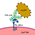
 Diagram of the functioning of a physical barrier ar.png 861 × 961; 336 кб
-
 Différentes modalités d'activation de la cellule NK.jpg 827 × 1192; 432 кб
-
 DQ Illustration.PNG 274 × 203; 7 кб
-
 DR beta 1 SEI topdown.JPG 244 × 164; 10 кб
-
 Eosinophil2.png 80 × 69; 8 кб
-
 Epitope.png 275 × 173; 4 кб
-
 Esempio extravasazione.svg 1968 × 998; 361 кб
-
 Familias de PRRs.png 1852 × 883; 497 кб
-
 Fattore C3 del Complemento Umano (2A73).png 1146 × 641; 2,8 Мб
-
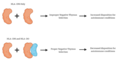
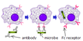 Fc receptor response.png 300 × 150; 13 кб
-
 Fc receptor schematic big.png 600 × 600; 10 кб
-
 FcAr.png 600 × 600; 14 кб
-
 Fcell-08-00677-g001.jpg 2474 × 1378; 377 кб
-
 Fpubh-08-00383-g003.jpg 1084 × 590; 513 кб
-
Gefahr durch chronische Entzündungskrankheiten.webm 43 с, 1920 × 1080; 43,37 Мб
-
 Genetic Background Causation.jpg 960 × 720; 49 кб
-
 HLA MHC Complex illustration.jpg 225 × 456; 17 кб
-
 HLA-DO Role.png 992 × 502; 60 кб
-
 HLAn(ro).png 230 × 435; 20 кб
-
Hoe versla jij elke dag zeven infecties en tien tumoren (1 5).webm 14 хв 59 с, 1920 × 1080; 160,96 Мб
-
 Homo sapiens CD8 molecule.png 640 × 480; 66 кб
-
How does your body beat a virus.webm 14 хв 29 с, 1920 × 1080; 331,73 Мб
-
 Human Paneth cells.JPG 2816 × 2112; 2,78 Мб
-
 Humeral Secondary Immune Response.png 5025 × 3137; 1,06 Мб
-
 Humoral Response Drawing.svg 512 × 384; 50 кб
-
 IgA antibody.tif 1280 × 720; 299 кб
-
 IL19 Crystal Structure.png 756 × 753; 205 кб
-
 ILC development 2 PNG.png 2283 × 2064; 678 кб
-
 Illu blood cell lineage (pt).png 480 × 350; 154 кб
-
 Immunantwort 1.png 1872 × 1368; 866 кб
-
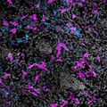 Immune Cells Surrounding Hair Follicles in Mouse Skin (7747026956).jpg 1200 × 1200; 261 кб
-
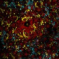 Immune Cells Surrounding Hair Follicles in Mouse Skin (7747051716).jpg 2100 × 2100; 685 кб
-
 Immune memory.png 1158 × 564; 507 кб
-
 Immune response Lymphocyte.svg 2170 × 1205; 489 кб
-
 Immune response of Lymphocytes.svg 2170 × 1205; 486 кб
-
 Immune response-ja.jpg 2271 × 1407; 253 кб
-
 Immune Response1.jpg 1024 × 768; 406 кб
-
 Immune.png 800 × 483; 68 кб
-
 Immunité passive Barrière intestinale.jpg 950 × 631; 133 кб
-
 Immunological Memory.png 5025 × 4579; 1,55 Мб
-
 InfiammazioneReazioneAcuta.png 1369 × 800; 748 кб
-
 Inflammatory response.jpg 1202 × 752; 78 кб
-
 Koemelkallegie-Eczeem in knieholte.jpg 480 × 640; 33 кб
-
 Lentinan2D.png 848 × 945; 17 кб
-
Liza Sumirat - Bagian 1 - Sistem Imun.wav 17 хв 26 с; 87,97 Мб
-
Liza Sumirat - Bagian 2 - Sistem Imun.wav 19 хв 37 с; 99,01 Мб
-
Liza Sumirat - Bagian 3 - Sistem Imun.wav 18 хв 50 с; 95,06 Мб
-
Liza Sumirat - Bagian 4 - Sistem Imun.wav 26 хв 21 с; 133,01 Мб
-
 Lymph Node Diagram Unlabeled.jpg 1812 × 1613; 231 кб
-
 Lymphocyte activation simple zh.png 612 × 358; 42 кб
-
 Lymphocyte activation simple-ca.png 612 × 358; 51 кб
-
 Lymphocyte activation simple-ja.png 2056 × 1193; 433 кб
-
 Lymphocyte activation simple.png 612 × 358; 52 кб
-
 Markers of non-self.jpg 570 × 427; 24 кб
-
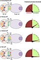
 Mechanisms of VSG switching2.png 1498 × 738; 504 кб
-
 Microchimerism.jpg 284 × 479; 18 кб
-
 Model of factorH-C3b complex.png 2999 × 2249; 1,88 Мб
-
 Mono-und-Polymere-zh.png 242 × 243; 20 кб
-
 Monoclonal antibodies4.jpg 1350 × 900; 624 кб
-
 Mouse IRG.png 640 × 480; 84 кб
-
 Mucosal immunity March 18.jpg 1517 × 960; 287 кб
-

-
 NFKB structure schematic.png 2359 × 943; 132 кб
-
 Normal T Cells (6830348943).jpg 106 × 160; 4 кб
-
 Normal T Cells (6830364101).jpg 160 × 109; 4 кб
-
 Opsonin cs.png 848 × 553; 92 кб
-
 PBB GE LCK 204890 s at tn.png 255 × 135; 541 байтаў
-
 PBB GE LCK 204891 s at tn.png 255 × 135; 550 байтаў
-
 PBB GE TLR4 221060 s at fs.png 732 × 530; 10 кб
-
 PBB Protein AIRE image.jpg 500 × 500; 16 кб
-
 Phagocytosis.JPG 599 × 410; 33 кб
-
 Phagocytosis.png 2352 × 1611; 101 кб
-
 Phagocytosis2.png 932 × 655; 101 кб
-
 PMAP-TLR.jpg 1000 × 402; 229 кб
-
 Primary immune response 1 ar.png 1872 × 1368; 857 кб
-
 Primary immune response 1.png 1872 × 1368; 864 кб
-

-
-
 Présentation de l'antigène.jpg 1000 × 623; 244 кб
-
 Putative mechaism of action of Human IRGM.jpg 688 × 511; 40 кб
-
 Relevant GO biological processes identified in the tear fluid.jpg 1200 × 976; 97 кб
-
 Reprogramming the immune system using ES cells..jpg 360 × 492; 61 кб
-
 Sample reaction norm graphic.jpg 500 × 439; 49 кб
-

-

-
 SCID joonis.tif 800 × 600; 1,83 Мб
-
 Selenium and anti-tumour immunity.jpg 2370 × 1936; 641 кб
-
 Selenium paradox.jpg 2362 × 1282; 290 кб
-
 Signal transduction pathways zh.png 1858 × 1364; 754 кб
-
 Signal transduction pathways.png 1858 × 1364; 709 кб
-
 Sistèma immunitari - Esquèma de la fagocitosi.png 495 × 1697; 159 кб
-

-
 Sistèma immunitari - Sistèma immunitari innat uman (esquèma generau).png 1016 × 1409; 322 кб
-
 Stat domain structure.png 600 × 86; 15 кб
-
 Síntesis d'icosanoides.jpg 693 × 611; 188 кб
-
 T-cell dependent b-cell act.jpg 720 × 540; 26 кб
-
 TCR complex.jpg 240 × 300; 10 кб
-
 Thyroid hormone pills - left T3 - right T4.jpg 1600 × 1200; 588 кб
-
 Tickover Initiation of the Alternative Complement Pathway.png 3984 × 2905; 947 кб
-
 TLR4.png 600 × 523; 271 кб
-
 Uw grafik immunsystem.jpg 800 × 599; 75 кб
-
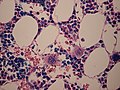 WVSOM Megakaryocytes.JPG 2816 × 2112; 2,03 Мб
-
 Zebra-fish phagocytes.jpg 1024 × 928; 367 кб
-
 Zellen des Immunsystems.jpg 2480 × 3508; 280 кб
-
 Макрофаги.jpg 230 × 230; 9 кб


.png)


.png)


.png)


.png)

.jpg)
.jpg)


.jpg)
.jpg)


..png)
.png)


