Category:Lymphatic system
Pereiti į navigaciją
Jump to search
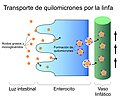
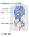
English: In mammals, including humans, the lymphatic system is composed of a network of thin vessels that branch, like blood vessels, into tissues throughout the body. Lymphatic vessels carry lymph, a colorless, watery fluid originating from interstitial fluid (fluid in the tissues) which is squeezed out of the blood vessels. The lymphatic system transports infection-fighting cells called lymphocytes, is involved in the removal of foreign matter and cell debris by phagocytes and is part of the body's immune system. One part of it also transports fats from the small intestine to the blood.
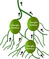
Lymphatic fluid gathers from the tissues and enters the valved lymphatic ducts. This 'lymph fluid' then passes back to the heart. On its way it passes through special nodular glands known as 'glands' or lymph nodes, which are concentrated in certain zones such as the back of the neck, the armpits and the groin. When a lymph node detects a possible threat passing into it in the lymph (such as a bacterium), it swells up. This is why lymph nodes swell in the region of an infected body part. Generalized lymphadenopathy (all the nodes of the body are swollen) can indicate systemic illness such infection or cancer. When generalized lymphadenopathy persists it is known as persistent generalized lymphadenopathy.
日本語: リンパ系
a part of the defense system (immune system) of vertebrate animals against pathogens  | |||||
| Įkelti mediją | |||||
| Tai yra |
| ||||
|---|---|---|---|---|---|
| Poklasis |
| ||||
| Yra dalis | |||||
| Susideda iš | |||||
| |||||
Subkategorijos
Šioje kategorijoje yra viena subkategorija.
Daugialypės terpės rinkmenos kategorijoje „Lymphatic system“
Šioje kategorijoje yra viena rinkmena.
-
 1Z3U.pdb1.jpg 550 × 550; 50 KiB
-
 3D medical animation TNM Staging System.jpg 1 920 × 1 080; 1 002 KiB
-
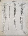
 Absorbing vessels of the human body, by William Cruikshank Wellcome L0013060.jpg 1 056 × 1 728; 1,02 MiB
-
 Anatomy of the lymphatic system.jpg 2 250 × 3 250; 651 KiB
-
 Circulacionlinfatica2.jpg 2 324 × 1 256; 307 KiB
-
 Circulatory system with labels.tif 2 250 × 3 250; 1,29 MiB
-
 Circulatory system.tif 2 250 × 3 250; 1,18 MiB
-

-

-
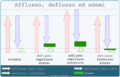 Edemi.png 800 × 512; 153 KiB
-
 Expansion of the lymphatic vasculature.jpg 2 250 × 3 250; 429 KiB
-
 Flusso linfatico.gif 78 × 500; 350 KiB
-
 G. Asellius, De lactibus sive lacteis Wellcome L0021461.jpg 1 155 × 1 674; 1,36 MiB
-
 Ganglioslinfaticosabdomen.jpg 1 280 × 1 024; 135 KiB
-
 Ganglioslinfaticosabdomenm.jpg 1 239 × 1 024; 182 KiB
-
 Gaspare Asellius, De lactibus sive lacteis venis Wellcome L0021944.jpg 1 121 × 1 714; 1,38 MiB
-
 Gray1064.png 400 × 474; 49 KiB
-
 Gray1191 zh.png 401 × 300; 80 KiB
-
 Gray1191.png 401 × 300; 34 KiB
-
 Homa limfo 001 1.jpg 435 × 1 284; 451 KiB
-
 Human embryo Recent findings about tissue-specific lymphatic progenitor cells..jpg 1 380 × 912; 289 KiB
-

-

-

-
 Insights from human genetics studies of primary lymphoedema.jpg 2 250 × 3 250; 483 KiB
-
 IPK.JPG 1 536 × 2 048; 708 KiB
-
 Keimzentrum.jpg 524 × 422; 62 KiB
-
 LEC specification in the cardinal vein.jpg 2 250 × 3 250; 301 KiB
-
 Logistica Perfusione.png 543 × 800; 253 KiB
-
 Lymfe-organen beenvis.svg 800 × 400; 454 KiB
-
 Lymph -.jpg 3 115 × 2 114; 1,15 MiB
-
 Lymphatics; Four figures showing the lymphatic system in the Wellcome V0007946ER.jpg 1 242 × 1 611; 1,29 MiB
-
 LymphaticSystem3.jpg 1 481 × 1 091; 170 KiB
-
 Lymphknoten (Schwein).jpg 2 229 × 1 516; 1,61 MiB
-
 Lymphocyte activation simple-es.png 612 × 358; 42 KiB
-
 Mandel entzuendung02.jpg 409 × 172; 60 KiB
-
 Maturation of the lymphatic system.jpg 4 000 × 2 250; 749 KiB
-
 MPP to DN3 dual myeloid and lymphoid.jpg 984 × 656; 200 KiB
-
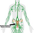 Módulos linfáticos iliacos internos.jpg 856 × 800; 89 KiB
-
 New Mixed Myeloid-Lymphoid Progenitor Tree(RCCH) Grayscale.jpg 1 268 × 587; 178 KiB
-
 Organos-linfoides.png 952 × 1 476; 611 KiB
-
 Perfusione.png 800 × 562; 250 KiB
-
 Pompe cardio - vascolari.png 478 × 800; 307 KiB
-
 Pompe venosa - linfatiche.png 600 × 552; 221 KiB
-
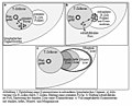
 Pressioni idrostatiche e osmotiche in capillari.png 800 × 432; 208 KiB
-
 Quilolinfa.jpg 5 144 × 4 150; 1,88 MiB
-
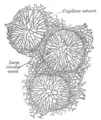
 Redlinfatica.jpg 2 428 × 2 946; 770 KiB
-
 Scambio di liquidi tra capillari e interstizio.png 800 × 421; 322 KiB
-
 Scambio sostanze capillari tessuti.png 812 × 800; 626 KiB
-
 Sistemalinfatico3.jpg 810 × 1 024; 181 KiB
-
 St Georges classification algorithm for primary lymphedema.jpg 3 551 × 2 523; 3,66 MiB
-
 St Georges Classification Algorithm.jpg 3 551 × 2 523; 1,95 MiB
-

-
-
 The Lacteals from Aselli. Wellcome M0010426.jpg 2 571 × 4 210; 4,45 MiB
-
 TJK Example lymphatics.jpg 512 × 512; 58 KiB
-
 Vessels and glands of the lymphatic system; seven figures, i Wellcome V0008037.jpg 2 948 × 2 380; 3,73 MiB
-
 БСЭ1. Лимфатическая система 1.jpg 221 × 63; 8 KiB
-
 БСЭ1. Лимфатическая система 2.jpg 308 × 578; 105 KiB
-
 БСЭ1. Лимфатическая система 3.jpg 308 × 578; 48 KiB
-
 БСЭ1. Лимфатическая система 4.jpg 215 × 352; 33 KiB


.jpg)


_Grayscale.jpg)


_(14596258808).jpg)
_(14769792951).jpg)


