Category:Mitosis
পরিভ্রমণে চলুন
অনুসন্ধানে চলুন
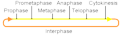
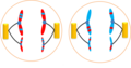
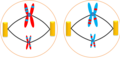
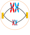
উন্নত শ্রেণীর প্রাণী ও উদ্ভিদের দেহকোষ বিভাজন প্রক্রিয়া  | |||||
| মিডিয়া আপলোড করুন | |||||
| নিদর্শন | |||||
|---|---|---|---|---|---|
| যার উপশ্রেণী | |||||
| এর অংশ |
| ||||
| এর ভিন্নরূপ | |||||
| |||||
উপবিষয়শ্রেণীসমূহ
এই বিষয়শ্রেণীতে অন্তর্ভুক্ত মোট ২০টি উপবিষয়শ্রেণীর মধ্যে ২০টি উপবিষয়শ্রেণী নিচে দেখানো হয়েছে।
A
C
- Cyclin B1 (7 F)
- Cyclin-dependent kinase 1 (11 F)
D
- Mitotic cells stained with DAPI (25 F)
M
- Mitotic signaling (9 F)
P
- Preprophase band (9 F)
- Prometaphase (8 F)
S
T
"Mitosis" বিষয়শ্রেণীতে অন্তর্ভুক্ত মিডিয়া ফাইলগুলি
এই বিষয়শ্রেণীতে অন্তর্ভুক্ত মোট ১৫৪টি পাতার মধ্যে ১৫৪টি পাতা নিচে দেখানো হলো।
-
 0300 Flourescence Stained.jpg ৬৭৫ × ৬৪৫; ২২৫ কিলোবাইট
-
 5 Makhluk Mitologi yang diyakini Dunia.pdf ১,৫০০ × ৮৪৩, ৭টি পাতা; ১.৮৮ মেগাবাইট
-
A-Role-for-the-Chaperone-Complex-BAG3-HSPB8-in-Actin-Dynamics-Spindle-Orientation-and-Proper-pgen.1005582.s007.ogv ৩.০ সে, ৫৫৬ × ৬৩৬; ২৭৪ কিলোবাইট
-
 Anaphase during Mitosis.svg ৫১২ × ৩৯৪; ৩২ কিলোবাইট
-
 Attivazione MPF (semplice).png ১,০৬৭ × ৪৪৯; ১৫ কিলোবাইট
-
 Cell cycle (5 stages of mitotic cell life).jpg ২,২০০ × ৫০০; ৪৫৭ কিলোবাইট
-
 Cell division according to A. Schneider (1873).png ১,২৬২ × ৩৫৭; ৮৩ কিলোবাইট
-
 Cell division according to E. Russov (1872).png ১,০৯০ × ৭৮৭; ৯৮ কিলোবাইট
-
 Cell division according to I. D. Chistyakov (1874).png ৫২০ × ৫৬৯; ৫২ কিলোবাইট
-
 Cell division according to W. Flemming (1882).png ১,২৮৬ × ১,৬৫২; ৪১৪ কিলোবাইট
-
 Cell division process.jpg ৭৬৬ × ৫৫৬; ৩০ কিলোবাইট
-
 Cell polarity.jpg ১৮১ × ১৬০; ৭ কিলোবাইট
-
 Cell proliferation.jpg ১,৫৫৭ × ৮৭৬; ৯৬ কিলোবাইট
-
 Cervical AIS, ThinPrep.jpg ৬৩৪ × ৬৪৩; ২১২ কিলোবাইট
-
 Chromatin bridge stained using DAPI 1.tiff ১,৩৯২ × ১,০৪০; ৪.১৪ মেগাবাইট
-
 Chromosomes in mitosis and meiosis.png ৩৯৬ × ৩৭৩; ১৭ কিলোবাইট
-
 Chromosomes2.jpg ২৫৬ × ২৫৬; ৪১ কিলোবাইট
-
 Cleavage Furrow Regression.svg ৫৩৬ × ৪৬১; ১১ কিলোবাইট
-
 Cleavage Furrow Regression.tif ৫৭২ × ৪৯২; ১৯ কিলোবাইট
-
 Com mROS contribueix a la senyalització mitogènica..gif ৪৪১ × ৫২২; ৭৮৭ কিলোবাইট
-
 Complete network.jpg ১,৩৯১ × ৮৬৭; ৯৮ কিলোবাইট
-
 Condensation and resolution of human sister chromatids in early mitosis.svg ৫১২ × ৫৮৫; ৯৩৪ কিলোবাইট
-
 CONDENSING CHROMOSOMES 2.jpg ১,৯৩৫ × ৫৪৯; ৭৮৫ কিলোবাইট
-
 Cormophyta alternancia-de-generaciones.png ১৯২ × ১৯২; ৯ কিলোবাইট
-
 Critique of the Theory of Evolution Fig 046.jpg ৫১০ × ৮২০; ৯৪ কিলোবাইট
-
 Cuerpo Flemming Asimetrica.jpg ৬৮০ × ৯০০; ৯২ কিলোবাইট
-
 Cuerpo Flemming Simetrico.jpg ৬৯২ × ৯০০; ৯৫ কিলোবাইট
-
 Cuerpo Flemming Ubicacion.png ৫৯২ × ৪৩৪; ২৭৬ কিলোবাইট
-
 Cuerpo intermedio Flemming.png ১,৯১২ × ৫৬০; ৪৩৩ কিলোবাইট
-
 Cytokinesis.png ২৮০ × ৩০২; ৪ কিলোবাইট
-
 Diagramatic illustration of the successive stages of mitosis (22699945268).jpg ১,৮৯০ × ২,৩৮৫; ১.২৯ মেগাবাইট
-
 Difference between Chromosome and Chromatid.png ১,৫২৮ × ৮৫০; ৫১ কিলোবাইট
-
 Difference between chromosomes and chromatids.png ১,৫১৪ × ৮৩৮; ১১০ কিলোবাইট
-
 Diploid Cell.png ১,৪২৪ × ৭৯২; ১২১ কিলোবাইট
-
 Division cellule.jpg ৪০০ × ১৪৭; ৬৯ কিলোবাইট
-
 DNA Replication in Diploid Cell.png ১,৪২৫ × ৭৯৯; ১২৫ কিলোবাইট
-
 EB1911 Cytology - heterotypical mitosis.jpg ৭৬০ × ২৭৭; ৬২ কিলোবাইট
-
 EB1911 Cytology - maturation divisions (1).jpg ৮১৩ × ৬৫৫; ১৭১ কিলোবাইট
-
 EB1911 Cytology - nuclear division.jpg ৭৬৬ × ৯৩৫; ২৭৪ কিলোবাইট
-
 EB1911 Cytology - preparation for mitosis (2).jpg ৮৯১ × ৪২১; ৯৯ কিলোবাইট
-
 EB1911 Cytology - preparation for mitosis.jpg ৭৯৩ × ২১১; ৫২ কিলোবাইট
-
 EB1911 Rhizopoda - Bud-fission of Euglypha alveolata.jpg ১,০৮৬ × ৭৩৮; ৩০৭ কিলোবাইট
-
 Embryo in flower.png ৩,০০০ × ৩,০০৬; ২.৯৭ মেগাবাইট
-
 Figure 04.jpg ৭০০ × ৯৫৫; ১০৪ কিলোবাইট
-
 Figure 05.jpg ৮০০ × ৬০৫; ৬৫ কিলোবাইট
-
 Figure 06.jpg ৪০০ × ১,০২৫; ৫৬ কিলোবাইট
-
 Figure 10 02 04.jpg ৫৪৪ × ৫৪৪; ১৭৩ কিলোবাইট
-
 Fluxograma - ativação da síntese de DNA por meio da ativação da pRb.jpg ৭৬২ × ৮৩৮; ৪৪ কিলোবাইট
-
 Fluxograma - atuação da pRb.jpg ৮৯৬ × ৮৭৩; ৭৯ কিলোবাইট
-
 Friedrich Reinke's medical school graduation document.jpg ১,৫৭৫ × ১,৯৫৯; ৩৩১ কিলোবাইট
-
 Gray2.png ৩৭৬ × ৬০০; ১৯ কিলোবাইট
-
 Gyhhhh.jpg ৭৮৮ × ৩৮৯; ৩২ কিলোবাইট
-

-
 Interphase cycle- Mitosis and Meiosis.png ১,৪১৬ × ৮১৭; ২৮৪ কিলোবাইট
-
 Interphase mitosis.png ৫০৮ × ১৪৯; ২ কিলোবাইট
-
 Irreversible Bistable Switch in Mitotic Exit.jpg ৯৬০ × ৬৩৩; ৫৪ কিলোবাইট
-
 MajorEventsInMitosis.jpg ৪৬০ × ১৬৭; ১৯ কিলোবাইট
-
 Malignant spindle cell neoplasm showing mitotic figures 40X.jpg ৯৯৭ × ৭৪৯; ২৮৭ কিলোবাইট
-
 Malignant spindle cell neoplasm showing mitotic figures.jpg ৯৯৭ × ৭৪৯; ২৭৩ কিলোবাইট
-
 Meiosis 1- Anaphase 1.png ৯৯৫ × ৭২৮; ৭৭ কিলোবাইট
-
 Meiosis 1- Cytokinesis.png ১,৬৬১ × ৮০৩; ১১০ কিলোবাইট
-
 Meiosis 1- Prophase 1.png ৯৯৮ × ৭১৩; ৬৩ কিলোবাইট
-
 Meiosis 1- Telophase 1.png ৬২৯ × ৭৩৩; ৫০ কিলোবাইট
-
 Meiosis 2- Anaphase 2.png ১,৩৭২ × ৭২৫; ১০২ কিলোবাইট
-
 Meiosis 2- Cytokinesis (2).png ১,৩২২ × ৭৭৭; ৯২ কিলোবাইট
-
 Meiosis 2- Cytokinesis.png ১,৩২২ × ৭৫৮; ৮৩ কিলোবাইট
-
 Meiosis 2- Metaphase 2.png ১,৩৭০ × ৭২৬; ১১৫ কিলোবাইট
-
 Meiosis 2- Prophase 2.png ১,৬৬১ × ৭২৫; ৬৭ কিলোবাইট
-
 Meiosis 2- Telophase 2.png ১,৩৭২ × ৭২৫; ৯৫ কিলোবাইট
-
 Meiosis- Metaphase 1.png ৬৩০ × ৭২৭; ৬৪ কিলোবাইট
-
 Meiosis1- Cytokinesis.png ১,৩৭২ × ৭৭৫; ৮১ কিলোবাইট
-
 Metaphase during Mitosis.svg ৫১২ × ৪৩১; ৪৯ কিলোবাইট
-
 Mitoos.jpg ১,০২২ × ৩৭২; ৫৮ কিলোবাইট
-
 Mitoosi1.jpg ৯৭ × ৫৫; ৩ কিলোবাইট
-
 Mitoosi2.jpg ১০৫ × ৭০; ৪ কিলোবাইট
-
 Mitoosi3.jpg ২৩০ × ৭৫; ৫ কিলোবাইট
-
 Mitos delar kromosomerna i en cellkärna..png ১,০২৩ × ৩৭২; ১২৭ কিলোবাইট
-
 Mitose colchicine fr.svg ৮০০ × ২৬৩; ১৩৫ কিলোবাইট
-
 Mitose.gif ৩৫৯ × ৫০১; ৪ কিলোবাইট
-
 Mitose.JPG ৬৯১ × ৬০০; ৪৮ কিলোবাইট
-
 Mitosiaren faseak - eu.svg ২,৩৬১ × ৪০৯; ১.১৬ মেগাবাইট
-
 Mitosis (1).jpg ১,৮০০ × ২,২৫৫; ৯২৯ কিলোবাইট
-
 Mitosis (13083175463).jpg ৫৯৫ × ৮৪২; ৬১ কিলোবাইট
-
 Mitosis Animation.gif ১,২০০ × ৬৭৫; ৩২৪ কিলোবাইট
-
 Mitosis by Elspeth.jpg ১,৪৪০ × ১,১৭৫; ২৭২ কিলোবাইট
-
 MITOSIS cells secuencie-es.jpg ১,০০০ × ১৪৯; ৪৯ কিলোবাইট
-
 Mitosis cells sequence English.svg ৭৭৪ × ১১৫; ৪৯৪ কিলোবাইট
-
 Mitosis classification.png ৬৮৩ × ৭৩৪; ১৩৬ কিলোবাইট
-
 Mitosis cycle.jpg ২,৪৬৪ × ২,০৫৬; ২.৮৭ মেগাবাইট
-
 Mitosis In A Lymphoma Cell.jpg ৭১৫ × ৬৬৭; ২৮১ কিলোবাইট
-
 Mitosis Mesenchymal Stem Cells.gif ৩০০ × ২৫৩; ২.৯৪ মেগাবাইট
-
 Mitosis of onion cells.jpg ৩,০২৪ × ৪,০৩২; ২.৪ মেগাবাইট
-
 Mitosis process.gif ৬০০ × ৪০০; ১২০ কিলোবাইট
-
 Mitosis schematic diagram-es.svg ৮৩৩ × ৭২৩; ১৯০ কিলোবাইট
-
 Mitosis Stages.JPG ২,৫৯২ × ১,৯৩৬; ২.২১ মেগাবাইট
-
 Mitosis stages.jpg ১,৫৬৪ × ১,৫৬৪; ১.২৩ মেগাবাইট
-
 Mitosis vs Meiosis Daughter Cells.png ১,২৫৪ × ৬৫৩; ১৫৬ কিলোবাইট
-
 Mitosis- Anaphase.png ৬২৫ × ৭৫১; ৫৪ কিলোবাইট
-
 Mitosis- Cytokinesis (1).png ১,৩৬৯ × ৬৬৩; ৮৪ কিলোবাইট
-
 Mitosis- Cytokinesis.png ১,৩৭২ × ৭৫৬; ৬৯ কিলোবাইট
-
 Mitosis- Metaphase.png ৬৪৬ × ৭৫৮; ৭১ কিলোবাইট
-
 Mitosis- Prophase.png ১,৪৯১ × ৮৩১; ১৬২ কিলোবাইট
-
 Mitosis- Telophase.png ৬২৫ × ৭৫৩; ৩৬ কিলোবাইট
-
 Mitosis-AscarisEggcs400x2.jpg ১,০২৪ × ৭৬৮; ১৪০ কিলোবাইট
-
 Mitosis.jpg ১৫৮ × ১৪২; ২৪ কিলোবাইট
-
 Mitosis.png ৫৭৬ × ৫২৮; ৮০ কিলোবাইট
-
 MitosisAndMeiosis de.png ১,২৭৪ × ১,৮০০; ২৩ কিলোবাইট
-
 MitosisAndMeiosis en.png ১,২৭৪ × ১,৮০০; ২৩ কিলোবাইট
-
 Mitotic Catastrophe Diagram 2.png ২,৯৫৮ × ১,৬৬৪; ১৯২ কিলোবাইট
-
 Mitotic Catastrophe Diagram version 2.png ২,৯৫৮ × ১,৬৬৪; ১৮৬ কিলোবাইট
-
 Mitotic Catastrophe Diagram.png ১,৭০৪ × ৯৬২; ৯৯ কিলোবাইট
-
 Mitotic checkpoint vertebrates.png ৬৮০ × ৫০১; ১৮ কিলোবাইট
-
 Mitotic LLC-PK1 cells, fluorescence microscopy (23700644352).jpg ১,৮০০ × ১,২০০; ১.৬ মেগাবাইট
-
 Network picture.png ৩৪৬ × ২৫৯; ৫৬ কিলোবাইট
-
 Neuroblast cell division - 486169.fig.002a.jpg ৬০০ × ২০৭; ১০ কিলোবাইট
-
 Nondisjunction in Mitosis.jpg ৪৭৯ × ৪৭৩; ৮৯ কিলোবাইট
-
 Normal and multipolar mitosis.tif ১,০২৪ × ১,০২৪; ৩.১৪ মেগাবাইট
-
 Nuclear envelope breakdown and reassembly in mitosis.jpg ১,২০০ × ৭৯০; ১৪২ কিলোবাইট
-
 Onion cells under a microscope.jpg ২,২৬৮ × ৪,০৩২; ১.৫২ মেগাবাইট
-
 Onion root cells.png ৯৬৫ × ৪৮২; ১,০১২ কিলোবাইট
-
 Pair-pull-part mitosis and meiosis.png ১,০২১ × ৭১৬; ২৯৫ কিলোবাইট
-
 Prophase diagram.svg ৫১২ × ৪০৭; ৪৬ কিলোবাইট
-
 PSM V71 D104 Cells from the mouth of the salamander to show mitosis.png ১,৩৮৭ × ২,০৬০; ৪২১ কিলোবাইট
-
 Remak cell1.jpg ২৫৬ × ১৯২; ১১ কিলোবাইট
-
RPE Cell Taxol treatment - mitotic catastrophe.webm ৮.৯ সে, ৬৫৪ × ৪৮০; ১.৮ মেগাবাইট
-
 RPE cell that has been treated with taxol and undergone mitotic catastrophe.png ২৫৬ × ৩০৪; ১২৪ কিলোবাইট
-
 Securin phosphorylation sites2.jpg ৪০৭ × ১৯৮; ৬২ কিলোবাইট
-
 Securin-separase conserved.png ৩৪৫ × ২৫৮; ৭৭ কিলোবাইট
-
 Simplified network controls mitotic exit.jpg ৯৬০ × ৬৩৩; ৫৫ কিলোবাইট
-
 Spindle Assembly Checkpoint.png ১,৯১৬ × ১,০৫১; ৩০৬ কিলোবাইট
-
 Spindle checkpoint vertebrates - en.png ৯৬০ × ৭২০; ৭৫ কিলোবাইট
-
 Stages of ciliate conjugation.svg ১,১৪৬ × ৮৬৩; ১১৬ কিলোবাইট
-
 Stages of early mitosis in a vertebrate cell (hy).svg ৫১২ × ২৩৯; ৬৯ কিলোবাইট
-
 Stages of early mitosis in a vertebrate cell with micrographs of chromatids.svg ৫১২ × ১,১৭০; ৪৫৮ কিলোবাইট
-
 Stages of early mitosis in a vertebrate cell.svg ৫১২ × ১,৪০৮; ১৫৬ কিলোবাইট
-
 Stages of late M phase in a vertebrate cell.svg ৫১২ × ১,৫৯৩; ২১১ কিলোবাইট
-
 StevensNM-StSp-1905-Pl-6-R12.jpg ১,৩৩৬ × ৫৭৮; ৮৭ কিলোবাইট
-
 StevensNM-StSp-1905-Pl-6-R34.jpg ১,৩৩৬ × ৫৮৬; ৯৮ কিলোবাইট
-
 StevensNM-StSp-1905-Pl-6-R56.jpg ১,৩৩৬ × ৬৩৭; ৯৬ কিলোবাইট
-
 StevensNM-StSp-1905-Pl-6-R70.jpg ১,৩৩৬ × ৩২১; ৫৪ কিলোবাইট
-
 Telophase during Mitosis.svg ৫১২ × ৩৮৪; ৫৫ কিলোবাইট
-
 The events of the eukaryotic cell cycle.svg ৫১২ × ২৫৬; ১.২৮ মেগাবাইট
-
 The twin brothers.tif ১০,৮৩৬ × ১০,০১১; ২১.৩১ মেগাবাইট
-
 Three cell growth types.png ৪৯৯ × ৫০৭; ১৭০ কিলোবাইট
-
 Trije tipi celične delitve.svg ৬৯৪ × ৭০০; ২৩৫ কিলোবাইট
-
 Tripolar Mitosis - breast carcinoma.jpg ২,০৪৮ × ১,৫৩৬; ১.৪১ মেগাবাইট
-
 Tripolar Mitosis - bronchial wash.jpg ১,২৩৮ × ৯২১; ৩৬৫ কিলোবাইট
-
 Twelve sketches illustrating all successive stages of mitosis (23129542051).jpg ১,৯২৫ × ১,৭৭২; ১.২৬ মেগাবাইট
-
 Types of mitosis.png ৬৬৪ × ৪৪৮; ১২১ কিলোবাইট
-
 Whole Genome Doubled Instability.png ৪,৪৩১ × ১,৩১৯; ৬৬৭ কিলোবাইট
-
 Wilson1900Fig1.jpg ১,৩৮৮ × ১,২০১; ২৯৬ কিলোবাইট
-
 Zellsubstanz-Kern-Kerntheilung.jpg ৩০৬ × ৪৩৪; ৩৬ কিলোবাইট
-
 Événements majeurs de la Mitose es.png ৪৬০ × ১৬৭; ৫৪ কিলোবাইট
-
 Митоза (анимација).gif ৮৬৫ × ৫৭৯; ৩.০১ মেগাবাইট


.png)
.png)
.png)
.png)


.jpg)


.jpg)
.jpg)


.png)


.jpg)
.jpg)


.png)


.jpg)


.svg)


.jpg)


.gif)
.jpg)
.png)