Category:Mitosis
Jump to navigation
Jump to search
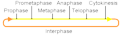
progression through the phases of the mitotic cell cycle, the most common eukaryotic cell cycle, which canonically comprises four successive phases called G1, S, G2, and M and includes replication of genome and subsequent segregation of chromosomes  | |||||
| Upload media | |||||
| Instance of | |||||
|---|---|---|---|---|---|
| Subclass of | |||||
| Has part(s) |
| ||||
| Different from | |||||
| |||||
Subcategories
This category has the following 20 subcategories, out of 20 total.
A
C
- Cyclin B1 (7 F)
- Cyclin-dependent kinase 1 (11 F)
D
- Mitotic cells stained with DAPI (25 F)
M
- Mitotic signaling (9 F)
P
- Preprophase band (9 F)
- Prometaphase (8 F)
S
T
Media in category "Mitosis"
The following 155 files are in this category, out of 155 total.
-
 0300 Flourescence Stained.jpg 675 × 645; 225 KB
-
 5 Makhluk Mitologi yang diyakini Dunia.pdf 1,500 × 843, 7 pages; 1.88 MB
-
-
 Anaphase during Mitosis.svg 512 × 394; 32 KB
-
 Attivazione MPF (semplice).png 1,067 × 449; 15 KB
-
 Cell cycle (5 stages of mitotic cell life).jpg 2,200 × 500; 457 KB
-
 Cell division according to A. Schneider (1873).png 1,262 × 357; 83 KB
-
 Cell division according to E. Russov (1872).png 1,090 × 787; 98 KB
-
 Cell division according to I. D. Chistyakov (1874).png 520 × 569; 52 KB
-
 Cell division according to W. Flemming (1882).png 1,286 × 1,652; 414 KB
-
 Cell division process.jpg 766 × 556; 30 KB
-
 Cell polarity.jpg 181 × 160; 7 KB
-
 Cell proliferation.jpg 1,557 × 876; 96 KB
-
 Cervical AIS, ThinPrep.jpg 634 × 643; 212 KB
-
 Chromatin bridge stained using DAPI 1.tiff 1,392 × 1,040; 4.14 MB
-
 Chromosomes in mitosis and meiosis.png 396 × 373; 17 KB
-
 Chromosomes2.jpg 256 × 256; 41 KB
-
 Cleavage Furrow Regression.svg 536 × 461; 11 KB
-
 Cleavage Furrow Regression.tif 572 × 492; 19 KB
-
 Com mROS contribueix a la senyalització mitogènica..gif 441 × 522; 787 KB
-
 Complete network.jpg 1,391 × 867; 98 KB
-

-
 CONDENSING CHROMOSOMES 2.jpg 1,935 × 549; 785 KB
-
 Cormophyta alternancia-de-generaciones.png 192 × 192; 9 KB
-
 Critique of the Theory of Evolution Fig 046.jpg 510 × 820; 94 KB
-
 Cuerpo Flemming Asimetrica.jpg 680 × 900; 92 KB
-
 Cuerpo Flemming Simetrico.jpg 692 × 900; 95 KB
-
 Cuerpo Flemming Ubicacion.png 592 × 434; 276 KB
-
 Cuerpo intermedio Flemming.png 1,912 × 560; 433 KB
-
 Cytokinesis.png 280 × 302; 4 KB
-
 Diagramatic illustration of the successive stages of mitosis (22699945268).jpg 1,890 × 2,385; 1.29 MB
-
 Difference between Chromosome and Chromatid.png 1,528 × 850; 51 KB
-
 Difference between chromosomes and chromatids.png 1,514 × 838; 110 KB
-
 Diploid Cell.png 1,424 × 792; 121 KB
-
 Division cellule.jpg 400 × 147; 69 KB
-
 DNA Replication in Diploid Cell.png 1,425 × 799; 125 KB
-
 EB1911 Cytology - heterotypical mitosis.jpg 760 × 277; 62 KB
-
 EB1911 Cytology - maturation divisions (1).jpg 813 × 655; 171 KB
-
 EB1911 Cytology - nuclear division.jpg 766 × 935; 274 KB
-
 EB1911 Cytology - preparation for mitosis (2).jpg 891 × 421; 99 KB
-
 EB1911 Cytology - preparation for mitosis.jpg 793 × 211; 52 KB
-
 EB1911 Rhizopoda - Bud-fission of Euglypha alveolata.jpg 1,086 × 738; 307 KB
-
 Embryo in flower.png 3,000 × 3,006; 2.97 MB
-
 Figure 04.jpg 700 × 955; 104 KB
-
 Figure 05.jpg 800 × 605; 65 KB
-
 Figure 06.jpg 400 × 1,025; 56 KB
-
 Figure 10 02 04.jpg 544 × 544; 173 KB
-

-
 Fluxograma - atuação da pRb.jpg 896 × 873; 79 KB
-
 Friedrich Reinke's medical school graduation document.jpg 1,575 × 1,959; 331 KB
-
 Gray2.png 376 × 600; 19 KB
-
 Gyhhhh.jpg 788 × 389; 32 KB
-

-
 Interphase cycle- Mitosis and Meiosis.png 1,416 × 817; 284 KB
-
 Interphase mitosis.png 508 × 149; 2 KB
-
 Irreversible Bistable Switch in Mitotic Exit.jpg 960 × 633; 54 KB
-
 MajorEventsInMitosis.jpg 460 × 167; 19 KB
-
 Malignant spindle cell neoplasm showing mitotic figures 40X.jpg 997 × 749; 287 KB
-
 Malignant spindle cell neoplasm showing mitotic figures.jpg 997 × 749; 273 KB
-
 Meiosis 1- Anaphase 1.png 995 × 728; 77 KB
-
 Meiosis 1- Cytokinesis.png 1,661 × 803; 110 KB
-
 Meiosis 1- Prophase 1.png 998 × 713; 63 KB
-
 Meiosis 1- Telophase 1.png 629 × 733; 50 KB
-
 Meiosis 2- Anaphase 2.png 1,372 × 725; 102 KB
-
 Meiosis 2- Cytokinesis (2).png 1,322 × 777; 92 KB
-
 Meiosis 2- Cytokinesis.png 1,322 × 758; 83 KB
-
 Meiosis 2- Metaphase 2.png 1,370 × 726; 115 KB
-
 Meiosis 2- Prophase 2.png 1,661 × 725; 67 KB
-
 Meiosis 2- Telophase 2.png 1,372 × 725; 95 KB
-
 Meiosis- Metaphase 1.png 630 × 727; 64 KB
-
 Meiosis1- Cytokinesis.png 1,372 × 775; 81 KB
-
 Metaphase during Mitosis.svg 512 × 431; 49 KB
-
 Mitoos.jpg 1,022 × 372; 58 KB
-
 Mitoosi1.jpg 97 × 55; 3 KB
-
 Mitoosi2.jpg 105 × 70; 4 KB
-
 Mitoosi3.jpg 230 × 75; 5 KB
-
 Mitos delar kromosomerna i en cellkärna..png 1,023 × 372; 127 KB
-
 Mitose colchicine fr.svg 800 × 263; 135 KB
-
 Mitose.gif 359 × 501; 4 KB
-
 Mitose.JPG 691 × 600; 48 KB
-
 Mitosiaren faseak - eu.svg 2,361 × 409; 1.16 MB
-
 Mitosis (1).jpg 1,800 × 2,255; 929 KB
-
 Mitosis (13083175463).jpg 595 × 842; 61 KB
-
 Mitosis Animation.gif 1,200 × 675; 324 KB
-
 Mitosis by Elspeth.jpg 1,440 × 1,175; 272 KB
-
 MITOSIS cells secuencie-es.jpg 1,000 × 149; 49 KB
-
 Mitosis cells sequence English.svg 774 × 115; 494 KB
-
 Mitosis classification.png 683 × 734; 136 KB
-
 Mitosis cycle.jpg 2,464 × 2,056; 2.87 MB
-
 Mitosis In A Lymphoma Cell.jpg 715 × 667; 281 KB
-
 Mitosis Mesenchymal Stem Cells.gif 300 × 253; 2.94 MB
-
 Mitosis of onion cells.jpg 3,024 × 4,032; 2.4 MB
-
 Mitosis process.gif 600 × 400; 120 KB
-
 Mitosis schematic diagram-es.svg 833 × 723; 190 KB
-
 Mitosis Stages.JPG 2,592 × 1,936; 2.21 MB
-
 Mitosis stages.jpg 1,564 × 1,564; 1.23 MB
-
 Mitosis vs Meiosis Daughter Cells.png 1,254 × 653; 156 KB
-
 Mitosis- Anaphase.png 625 × 751; 54 KB
-
 Mitosis- Cytokinesis (1).png 1,369 × 663; 84 KB
-
 Mitosis- Cytokinesis.png 1,372 × 756; 69 KB
-
 Mitosis- Metaphase.png 646 × 758; 71 KB
-
 Mitosis- Prophase.png 1,491 × 831; 162 KB
-
 Mitosis- Telophase.png 625 × 753; 36 KB
-
 Mitosis-AscarisEggcs400x2.jpg 1,024 × 768; 140 KB
-
 Mitosis.jpg 158 × 142; 24 KB
-
 Mitosis.png 576 × 528; 80 KB
-
 MitosisAndMeiosis de.png 1,274 × 1,800; 23 KB
-
 MitosisAndMeiosis en.png 1,274 × 1,800; 23 KB
-
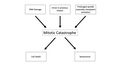 Mitotic Catastrophe Diagram 2.png 2,958 × 1,664; 192 KB
-
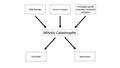 Mitotic Catastrophe Diagram version 2.png 2,958 × 1,664; 186 KB
-
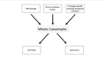 Mitotic Catastrophe Diagram.png 1,704 × 962; 99 KB
-
 Mitotic checkpoint vertebrates.png 680 × 501; 18 KB
-
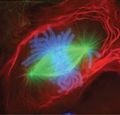
 Mitotic LLC-PK1 cells, fluorescence microscopy (23700644352).jpg 1,800 × 1,200; 1.6 MB
-
 Network picture.png 346 × 259; 56 KB
-
 Neuroblast cell division - 486169.fig.002a.jpg 600 × 207; 10 KB
-
 Nondisjunction in Mitosis.jpg 479 × 473; 89 KB
-
 Normal and multipolar mitosis.tif 1,024 × 1,024; 3.14 MB
-
 Nuclear envelope breakdown and reassembly in mitosis.jpg 1,200 × 790; 142 KB
-
 Onion cells under a microscope.jpg 2,268 × 4,032; 1.52 MB
-
 Onion root cells.png 965 × 482; 1,012 KB
-
 Pair-pull-part mitosis and meiosis.png 1,021 × 716; 295 KB
-

-
 Prophase diagram.svg 512 × 407; 46 KB
-
 PSM V71 D104 Cells from the mouth of the salamander to show mitosis.png 1,387 × 2,060; 421 KB
-
 Remak cell1.jpg 256 × 192; 11 KB
-
RPE Cell Taxol treatment - mitotic catastrophe.webm 8.9 s, 654 × 480; 1.8 MB
-

-
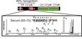 Securin phosphorylation sites2.jpg 407 × 198; 62 KB
-
 Securin-separase conserved.png 345 × 258; 77 KB
-
 Simplified network controls mitotic exit.jpg 960 × 633; 55 KB
-
 Spindle Assembly Checkpoint.png 1,916 × 1,051; 306 KB
-
 Spindle checkpoint vertebrates - en.png 960 × 720; 75 KB
-
 Stages of ciliate conjugation.svg 1,146 × 863; 116 KB
-
 Stages of early mitosis in a vertebrate cell (hy).svg 512 × 239; 69 KB
-
 Stages of early mitosis in a vertebrate cell with micrographs of chromatids.svg 512 × 1,170; 458 KB
-
 Stages of early mitosis in a vertebrate cell.svg 512 × 1,408; 156 KB
-
 Stages of late M phase in a vertebrate cell.svg 512 × 1,593; 211 KB
-
 StevensNM-StSp-1905-Pl-6-R12.jpg 1,336 × 578; 87 KB
-
 StevensNM-StSp-1905-Pl-6-R34.jpg 1,336 × 586; 98 KB
-
 StevensNM-StSp-1905-Pl-6-R56.jpg 1,336 × 637; 96 KB
-
 StevensNM-StSp-1905-Pl-6-R70.jpg 1,336 × 321; 54 KB
-
 Telophase during Mitosis.svg 512 × 384; 55 KB
-
 The events of the eukaryotic cell cycle.svg 512 × 256; 1.28 MB
-
 The twin brothers.tif 10,836 × 10,011; 21.31 MB
-
 Three cell growth types.png 499 × 507; 170 KB
-
 Trije tipi celične delitve.svg 694 × 700; 235 KB
-
 Tripolar Mitosis - breast carcinoma.jpg 2,048 × 1,536; 1.41 MB
-
 Tripolar Mitosis - bronchial wash.jpg 1,238 × 921; 365 KB
-
 Twelve sketches illustrating all successive stages of mitosis (23129542051).jpg 1,925 × 1,772; 1.26 MB
-
 Types of mitosis.png 664 × 448; 121 KB
-
 Whole Genome Doubled Instability.png 4,431 × 1,319; 667 KB
-
 Wilson1900Fig1.jpg 1,388 × 1,201; 296 KB
-
 Zellsubstanz-Kern-Kerntheilung.jpg 306 × 434; 36 KB
-
 Événements majeurs de la Mitose es.png 460 × 167; 54 KB
-
 Митоза (анимација).gif 865 × 579; 3.01 MB


.png)
.png)
.png)
.png)


.jpg)


.jpg)
.jpg)


.png)


.jpg)
.jpg)


.png)


.jpg)


..jpg)


.svg)


.jpg)


.gif)
.jpg)
.png)