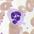
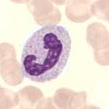
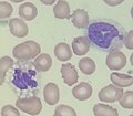
Category:Neutrophils
Перейти до навігації
Перейти до пошуку
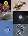
most abundant white blood cell, a type of granulocyte  | |||||
| Завантажити медіафайл | |||||
| Є одним із | |||||
|---|---|---|---|---|---|
| Є підкласом | |||||
| Частина від | |||||
| Не плутати з | |||||
| |||||
Підкатегорії
Показано 9 підкатегорій із 9.
- Videos of Neutrophils (120 F)
B
- Band neutrophils (16 F)
N
- Neutropenia (5 F)
- Neutrophil activation (7 F)
- Neutrophil cytoplasmic anomalies (16 F)
- Neutrophil infiltration (13 F)
- Neutrophil nuclear segmentation (22 F)
Файли в категорії «Neutrophils»
Показано 93 файли цієї категорії (із 93).
-
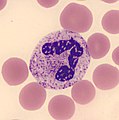
 20100825 023736 Neutrophil.jpg 750 × 750; 50 КБ
-
 Amoeba collage.jpg 2550 × 3300; 984 КБ
-

-
 ANCA ETHANOL AND FORMALIN.JPEG 2918 × 2189; 5,71 МБ
-
 Apoptotic neutrophil with nuclear fragmentation.jpg 110 × 101; 11 КБ
-
 Band form neutrophil in Wright stained PBS microscopy.jpg 3264 × 2448; 950 КБ
-
 Basics of neutrophils image cont.png 1142 × 902; 1015 КБ
-
 Basics of neutrophils image.png 1422 × 1056; 1,4 МБ
-
 Blausen 0676 Neutrophil (crop).png 1165 × 1130; 1,62 МБ
-
 Blausen 0676 Neutrophil.png 1500 × 1500; 1,34 МБ
-
 Blausen 0909 WhiteBloodCells.png 1600 × 1200; 1,23 МБ
-
 Blood-neutrophil1.jpg 829 × 745; 477 КБ
-
 Blood-neutrophil2.jpg 500 × 375; 214 КБ
-
 Blood-neutrophil3.jpg 500 × 375; 213 КБ
-
 Blood-neutrophil4.jpg 500 × 375; 218 КБ
-
 Bone marrow WBC.JPG 2272 × 1704; 1,72 МБ
-
 C anca.jpg 372 × 393; 144 КБ
-
 C ANCA.jpg 2048 × 1536; 2,77 МБ
-
 Cayado con granulación tóxica.jpg 1533 × 1533; 289 КБ
-
 Célula faríngea y neutrófilos con estreptococos grupo A en tinción Gram 400 x.jpg 3840 × 2160; 1,83 МБ
-
 Eosinophil and polymorphonuclear neutrophil.jpg 1280 × 960; 968 КБ
-

-
 G Neutrophile; G éosinophile.jpg 400 × 402; 55 КБ
-
 Green neutrophilic inclusions.jpg 489 × 503; 311 КБ
-
 Happy neutrophil.jpg 2016 × 1512; 145 КБ
-
 Hem1SegmNeutrophile.jpg 360 × 363; 19 КБ
-
 Hem1SegmNeutrophile2.jpg 360 × 363; 20 КБ
-
 Hem1SegmNeutrophile3.jpg 360 × 363; 16 КБ
-
 Hem1SegmNeutrophile4.jpg 360 × 363; 17 КБ
-
 Histopathology of neutrophil infiltration in myocardial infarction.jpg 1804 × 1218; 437 КБ
-
 Human Cell Groups distributed by Cell Count and by Aggregate Cell Mass.jpg 3162 × 2096; 1,08 МБ
-
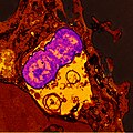
 Human neutrophil ingesting MRSA.jpg 1894 × 2501; 2,42 МБ
-
 Hypersegmented neutrophil - by Gabriel Caponetti,MD.jpg 435 × 380; 76 КБ
-
 Hypersegmented PMN.JPG 2272 × 1704; 1,11 МБ
-
-
 Lung biopsy showing lobar pneumonia 10X.jpg 862 × 649; 156 КБ
-
 Lung biopsy showing lobar pneumonia 40X.jpg 804 × 605; 109 КБ
-
 Margination of neutrophils in acute inflammation.png 724 × 540; 1012 КБ
-
 Margination of neutrophils.jpg 2048 × 1536; 515 КБ
-
 Migrace neutrofilů do místa infekce.jpg 912 × 658; 99 КБ
-
 Neutrofeeling amazin.jpg 2560 × 1920; 3,1 МБ
-
 Neutrofilo.JPG 3072 × 2304; 2,45 МБ
-
 Neutrophil (30104264763).jpg 1633 × 1830; 365 КБ
-
 Neutrophil (sketch).png 100 × 83; 6 КБ
-
 Neutrophil + monocyte (16076054013).jpg 747 × 747; 276 КБ
-
 Neutrophil 1.jpg 1280 × 720; 116 КБ
-
-
 Neutrophil in a blood smear.jpg 540 × 428; 51 КБ
-
 Neutrophil MRSA I.jpg 907 × 949; 679 КБ
-
 Neutrophil MRSA II.jpg 1064 × 806; 820 КБ
-
 Neutrophil with anthrax copy.jpg 2304 × 2403; 2,28 МБ
-
 Neutrophil with anthrax.jpg 2304 × 2403; 2,14 МБ
-
 Neutrophil.jpg 800 × 600; 82 КБ
-
 Neutrophil.png 80 × 79; 5 КБ
-
 Neutrophil2.jpg 290 × 261; 76 КБ
-
 Neutrophil3.tif 514 × 452; 681 КБ
-
 Neutrophiler Granulozyt.JPG 4000 × 3000; 4,45 МБ
-
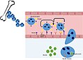
 NeutrophilerAktion.png 574 × 701; 126 КБ
-
 NeutrophilerAktion.svg 598 × 756; 102 КБ
-
 Neutrophils + monocyte (16694967012).jpg 858 × 858; 815 КБ
-
 Neutrophils -1.jpg 2048 × 1536; 1,16 МБ
-
 Neutrophils phagocytizing bacteria.jpg 700 × 463; 40 КБ
-
 Neutrophils with intracellular bacteria on peripheral blood smear.png 623 × 281; 291 КБ
-
 Neutrophils with segmented nuclei.jpg 2464 × 2056; 3,15 МБ
-
 Neutrophils-a type of leucocyte.jpg 2560 × 2048; 214 КБ
-
 Neutrophils-hue-gray.jpg 800 × 583; 174 КБ
-
 Neutrophils-hue-threshold-linear.jpg 800 × 583; 165 КБ
-
 Neutrophils-hue.jpg 800 × 583; 243 КБ
-
 Neutrophils.jpg 1455 × 1060; 201 КБ
-
 Neutrófilo (Neutrophil) (36583987456).jpg 1176 × 1344; 447 КБ
-
 Neutrófilo segmentado con granulación tóxica.jpg 1512 × 1512; 279 КБ
-
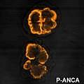 P anca.jpg 347 × 343; 129 КБ
-
 P ANCA.jpg 2048 × 1536; 1,96 МБ
-
 Parasite150072-fig1 Role of neutrophils in rodent amebic liver abscess.png 2667 × 2645; 722 КБ
-
 Parasite150072-fig2 Role of neutrophils in rodent amebic liver abscess.png 3693 × 3751; 1,36 МБ
-
 PBNeutrophil.jpg 150 × 150; 3 КБ
-
 PBStabkerniger.jpg 150 × 150; 2 КБ
-
 Pelger-Hüet Anomaly Overview.jpg 2920 × 2942; 1,13 МБ
-
 Pelgeroid Neutrophil (bilobate) (360621427).jpg 695 × 508; 282 КБ
-
 Pelgeroid Neutrophil (monolobate) (360621424).jpg 700 × 538; 287 КБ
-
 Promyelocyte.JPG 1024 × 768; 242 КБ
-
 Pyogenic meningitis.jpg 1200 × 1600; 335 КБ
-
 Seg-hemo.JPG 65 × 65; 2 КБ
-
 Segmented neutrophil (16508611940).jpg 956 × 956; 1018 КБ
-
 Segmented neutrophils.jpg 800 × 610; 382 КБ
-
 SEM blood cells.jpg 1800 × 2239; 1,33 МБ
-
 Toxic vacuolation 2.jpg 477 × 489; 310 КБ
-
 Toxic vacuolation.jpg 825 × 723; 370 КБ
-
 Vývoj neutrofilů v kostní dřeni.png 1523 × 334; 32 КБ
-
 WBC precursors.JPG 1024 × 768; 258 КБ
-
 נוטרופילים.tif 1280 × 1024; 1,29 МБ
-
 രക്തകോശങ്ങൾ.jpg 1800 × 2239; 1,38 МБ
-
 好中球の分化.png 1100 × 226; 41 КБ


.png)


_containing_ingested_Klebsiella_pneumoniae_(purple).jpg)


.jpg)
.png)
.jpg)
_Bacteria.jpg)


.jpg)


_(36583987456).jpg)


_(360621427).jpg)
_(360621424).jpg)


.jpg)


