Category:Ophthalmology
Vai alla navigazione
Vai alla ricerca

branca medica che si occupa dei disturbi della vista  | |||||
| Carica un file multimediale | |||||
| Istanza di | |||||
|---|---|---|---|---|---|
| Sottoclasse di |
| ||||
| Parte di |
| ||||
| |||||
 | |||||


Sottocategorie
Questa categoria contiene le 32 sottocategorie indicate di seguito, su un totale di 32.
.
A
B
D
E
- Ophthalmic events (2 F)
H
M
- Media from BMC Ophthalmology (12 F)
O
- Ocular dominance (12 F)
- Oculus (company) (5 F)
P
- Pupillary light reflex (10 F)
S
V
File nella categoria "Ophthalmology"
Questa categoria contiene 75 file, indicati di seguito, su un totale di 75.
-
 Should not oculists be more careful in prescribing colored glasses? (IA 101167335.nlm.nih.gov).pdf 629 × 989, 4 pagine; 153 KB
-
 A woman's head and neck; her eyes are closed and her mouth t Wellcome V0029467.jpg 2 400 × 2 945; 3,12 MB
-
 A woman's head and shoulders, her right eye is facing in a d Wellcome V0029482.jpg 2 450 × 2 897; 2,23 MB
-
 A woman's head and shoulders, her right eyelid is heavier th Wellcome V0029481.jpg 2 400 × 2 943; 2,8 MB
-
 A woman, head and shoulders, viewed from the front; her eyel Wellcome V0029454.jpg 2 350 × 2 991; 2,84 MB
-
 A woman, head and shoulders, viewed from the front; her eyel Wellcome V0029455.jpg 2 316 × 2 964; 2,34 MB
-
 ACC Anterior Chamber Cell Grading Scale.png 572 × 448; 186 KB
-
 Allergic shiner in pediatric patient.png 720 × 540; 644 KB
-
 ANALISI DELL'OCCHIO 1.jpg 3 330 × 2 079; 539 KB
-
-
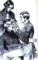 Anastasio Symphronio de Abreu Erlangen.jpg 563 × 889; 151 KB
-
 Anastasio Symphronio de Abreu.jpg 643 × 923; 69 KB
-
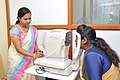 AR Examination.jpg 3 216 × 2 136; 2,85 MB
-
 Arzneibuch. Western Manuscript 990, page 83 Wellcome L0017448.jpg 1 232 × 1 558; 1,27 MB
-

-
 Gesichtsstörungen und Uterinleiden (electronic resource) (IA b21645267).pdf 902 × 1 500, 66 pagine; 2,8 MB
-

-
 Białystok 2023 132.jpg 3 000 × 4 000; 4,25 MB
-
 Caen Armenicus TEI.jpg 2 182 × 2 407; 711 KB
-
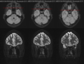 Case-3-mri.png 813 × 615; 368 KB
-
 Co01png.png 928 × 634; 619 KB
-
 Co03png.png 925 × 1 350; 2,24 MB
-
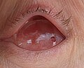 Conformer over eye implant after enucleation.jpg 951 × 769; 115 KB
-
 Cyclopentolate 1 percent Pupils.jpg 886 × 194; 120 KB
-
 EBO Signet.jpg 296 × 128; 9 KB
-
 Eye chart visual acuity simulator GIF.gif 1 092 × 728; 1,13 MB
-
 Eye treatment.jpg 4 000 × 6 000; 5,9 MB
-
 Fundus geographic atrophy.jpg 1 409 × 1 406; 132 KB
-
 Fundus photo Retina OD.jpg 15 997 × 15 989; 68,16 MB
-
 Fundus photograph Retina OS.jpg 15 997 × 15 989; 74,22 MB
-
 Goldmann Applanation Tonometer Labeled.png 457 × 815; 519 KB
-
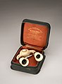 Ideal Sight Restorer, New York, United States, 1901-1930 Wellcome L0058540.jpg 2 832 × 3 844; 1,39 MB
-
 IN A MICROSCOPE.jpg 3 024 × 4 032; 814 KB
-
 Kamienica, mur., 1870 Płock, ul. Tumska 14.jpg 4 592 × 3 056; 5,54 MB
-
 Lanterntest Beyne Replikat.jpg 3 024 × 4 032; 2,7 MB
-
 Lens Cornea Transmission.png 1 655 × 985; 106 KB
-
 Lunghezze d'onda coni e bastoncelli retina.png 1 856 × 2 048; 626 KB
-
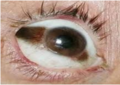
 Malignant growth springing from the eye of a woman Wellcome L0061858.jpg 4 552 × 5 224; 4,16 MB
-
-
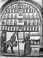 Manuscript Arzneibuch. Wellcome M0007369.jpg 2 800 × 3 845; 2,69 MB
-
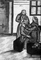 Manuscript Arzneibuch. Wellcome M0007411.jpg 2 700 × 3 966; 2,94 MB
-
 Mario Romano.jpg 1 024 × 614; 91 KB
-
 MEHRI Logo copy.jpg 1 768 × 812; 337 KB
-
 Monocular Lens Transmittance.png 3 050 × 1 804; 331 KB
-
 Near point, calculated from Duane's (1922) original curve.png 2 211 × 1 707; 77 KB
-
 Ocular OCT 3D View OD.gif 1 436 × 889; 5,01 MB
-
 Ocular OCT OD IR30 no overlay.jpg 1 536 × 1 536; 1,91 MB
-
 Ocular OCT OS IR30 overlay.jpg 768 × 768; 438 KB
-
 OD Ocular IROCT 30 ART no overlay.gif 1 008 × 496; 8,5 MB
-
 OD Ocular IROCT 30 ART ON no overlay.gif 1 008 × 496; 4,88 MB
-
 Offspring eye color.png 1 080 × 1 080; 496 KB
-

-
 OphthalmicSolutions.jpg 1 527 × 1 413; 235 KB
-
 OPN-recombination-chimeras.gif 1 577 × 602; 27 KB
-
 OPN-recombination-dichromacy.gif 1 577 × 602; 28 KB
-
 Optic Neuritis.png 1 257 × 476; 609 KB
-
 OS Ocular IROCT 30 ART ON no overlay.gif 1 008 × 496; 4,55 MB
-
 OS Ocular IROCT 30 ART overlay.gif 1 008 × 496; 8,07 MB
-
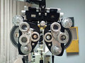 Outward-facing side of a phoropter (50987068248).png 3 100 × 2 325; 7,85 MB
-
 Placido's disc 2.jpg 3 456 × 4 608; 6,87 MB
-
 Placido's disc 3.jpg 3 456 × 4 608; 6,64 MB
-
 Placido's disc.jpg 3 456 × 4 608; 6,89 MB
-
 Rayleigh Match Anomaloscope.png 1 674 × 885; 36 KB
-
 Recommended Levels of Blue Light Exposure By Age.png 1 271 × 301; 270 KB
-
 Refining Innovation 160223-A-AP268-244.jpg 4 288 × 2 848; 5,37 MB
-
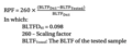 RPF Formula.png 386 × 149; 27 KB
-
 Sede Clínica Oftalmológica del Caribe.jpg 1 629 × 1 123; 278 KB
-
 STS-41 crewmembers conduct DSO 0474 Retinal Photography on OV-103's middeck.jpg 4 096 × 2 664; 2,26 MB
-
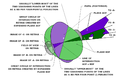 Test Fig..png 922 × 600; 68 KB
-
 The British Army in Italy 1944 NA13011.jpg 800 × 797; 88 KB
-
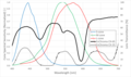 Transmittance EnChroma.png 3 051 × 1 804; 322 KB
-
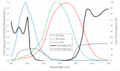 Transmittance Tinted Lenses.png 3 050 × 1 804; 319 KB
-
 Variantor transmittance.png 3 049 × 1 803; 286 KB
-
 Waren Tay (cropped).png 289 × 386; 61 KB
-
 Waren Tay.png 402 × 619; 258 KB


.png)


_original_curve.png)


.png)


.png)
