Category:Organelles
Salti al navigilo
Salti al serĉilo
organized cell-level biological structure of distinctive morphology and function | |||||
| Alŝuti plurmedion | |||||
| Estas |
| ||||
|---|---|---|---|---|---|
| Subaro de |
| ||||
| |||||
Subkategorioj
Ĉi tiu kategorio havas la 35 jenajn subkategoriojn, el 35 entute.
- Animations of organelles (1 D)
?
- Organelle shape (15 D)
A
- Apoptosomes (13 D)
B
- Birbeck granules (2 D)
C
E
F
G
H
- Haptonema (1 D)
J
- JUNQ and IPOD (5 D)
L
- Lipid droplets (83 D)
M
- Micronuclei (9 D)
- Microvesicles (11 D)
O
- Organelle biogenesis (10 D)
- Organelle interactions (7 D)
- Organelle size (5 D)
P
R
S
V
- Vacuole (11 D)
W
Dosieroj en kategorio “Organelles”
La jenaj 43 dosieroj estas en ĉi tiu kategorio, el 43 entute.
-

-

-
-

-

-

-
 AML-M5B, promonocyte, TEM.jpg 685 × 512; 309 KB
-
 Cell parts.png 492 × 259; 57 KB
-
 Celorganellen van een dierlijke cel.png 813 × 645; 451 KB
-
 Cilium Anderson.jpg 350 × 477; 53 KB
-
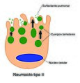 Cuerposlamelares.jpg 2 244 × 2 346; 1,12 MB
-
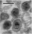 Electron micrograph of microvesicles.tiff 313 × 332; 111 KB
-
 Endomembrane system diagram no text nucleus.png 385 × 414; 84 KB
-
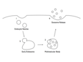 Exosome formation..tiff 1 024 × 768; 75 KB
-
 Exosome Formation.png 1 024 × 768; 65 KB
-
 Golgi secretions.png 432 × 301; 68 KB
-
 HeLa pspecks2 gl.jpg 283 × 298; 24 KB
-
 HeLa pspecks2.jpg 283 × 298; 46 KB
-
 HeuserAxo.jpg 374 × 500; 113 KB
-
 Hydrogenosomal activity.webp 946 × 490; 47 KB
-
 ICON for template of cell organelles.png 1 389 × 1 355; 1,58 MB
-
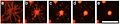
 Journal.pone.0031641.g003.png 902 × 830; 2,4 MB
-
 Lisosoma.png 670 × 854; 150 KB
-
 Loukoumasome.png 978 × 2 280; 1,35 MB
-
 Magnetosoma.png 241 × 149; 44 KB
-
 Magnetosome Chain Formation.png 566 × 308; 82 KB
-
 Magnetosome collapse.png 467 × 506; 159 KB
-
 Minor spliceosome.jpg 585 × 951; 170 KB
-
 Mitokondrid, mikrofilamendid ja tuumad rakkudes.jpg 5 648 × 3 566; 9,9 MB
-
 Nucleo-RER.jpg 1 000 × 1 185; 332 KB
-
 Parenthesome miguelferig.jpg 1 312 × 697; 60 KB
-

-
 Plant cell structure svg vacuole (id).jpg 649 × 475; 156 KB
-
 PZSL1889Plate05.png 1 793 × 2 916; 3,39 MB
-
 Retikulum stanica.png 500 × 540; 97 KB
-
 Ribosomi stanica.png 500 × 540; 82 KB
-
 Rol de les flipases en la biogènesi vesicular.jpg 430 × 201; 26 KB
-
 Trypanosomeglycosomes.jpeg 200 × 345; 29 KB
-
 Ubiquitylation.png 800 × 486; 115 KB
-
 Vacuole and nucleus in aloe vera cell.jpg 640 × 480; 216 KB
-
 Yeast tri-snRNP.jpg 2 763 × 2 011; 1,06 MB
-
 Zellen-chl-mito-pero.png 715 × 668; 564 KB
-
 سلول جانوری فارسی.jpg 624 × 417; 107 KB


.jpg)


