Category:Cell membrane
Jump to navigation
Jump to search
biological membrane that separates the interior of a cell from its outside environment  | |||||
| Upload media | |||||
| Instance of |
| ||||
|---|---|---|---|---|---|
| Subclass of |
| ||||
| Part of |
| ||||
| |||||
Subcategories
This category has the following 16 subcategories, out of 16 total.
Media in category "Cell membrane"
The following 200 files are in this category, out of 669 total.
(previous page) (next page)-
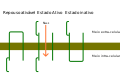
 20the-20plasma-20membrane.png 884 × 179; 13 KB
-
A-Role-for-the-Membrane-in-Regulating-Chlamydomonas-Flagellar-Length-pone.0053366.s004.ogv 12 s, 320 × 372; 2.61 MB
-
A-Role-for-the-Membrane-in-Regulating-Chlamydomonas-Flagellar-Length-pone.0053366.s005.ogv 12 s, 316 × 284; 8.83 MB
-
A-Role-for-the-Membrane-in-Regulating-Chlamydomonas-Flagellar-Length-pone.0053366.s007.ogv 12 s, 508 × 340; 5.2 MB
-
-
-
-
-
-
-
-
-
-
-
-
 Alpha Intercalated Cell Cartoon.jpg 777 × 604; 45 KB
-
 Asimmetria membrana.jpg 960 × 720; 106 KB
-
 Black lipid membrane.svg 709 × 590; 122 KB
-
 Canais.svg 610 × 387; 8 KB
-
 Cell membrane in Rheo leaf cell.jpg 3,120 × 1,440; 1.71 MB
-
 Cell Membrane.png 1,064 × 561; 207 KB
-
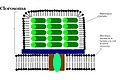 Clorosome.JPG 829 × 573; 61 KB
-
Control-of-Directed-Cell-Migration-In-Vivo-by-Membrane-to-Cortex-Attachment-pbio.1000544.s009.ogv 6.2 s, 264 × 264; 701 KB
-
Control-of-Directed-Cell-Migration-In-Vivo-by-Membrane-to-Cortex-Attachment-pbio.1000544.s010.ogv 6.2 s, 264 × 264; 388 KB
-
Control-of-Directed-Cell-Migration-In-Vivo-by-Membrane-to-Cortex-Attachment-pbio.1000544.s011.ogv 6.2 s, 264 × 264; 454 KB
-
Control-of-Directed-Cell-Migration-In-Vivo-by-Membrane-to-Cortex-Attachment-pbio.1000544.s012.ogv 7.6 s, 418 × 416; 1.51 MB
-
Control-of-Directed-Cell-Migration-In-Vivo-by-Membrane-to-Cortex-Attachment-pbio.1000544.s013.ogv 7.5 s, 419 × 416; 680 KB
-
Control-of-Directed-Cell-Migration-In-Vivo-by-Membrane-to-Cortex-Attachment-pbio.1000544.s014.ogv 6.5 s, 512 × 512; 532 KB
-
-
Control-of-Directed-Cell-Migration-In-Vivo-by-Membrane-to-Cortex-Attachment-pbio.1000544.s016.ogv 11 s, 1,024 × 529; 2.41 MB
-
Control-of-Directed-Cell-Migration-In-Vivo-by-Membrane-to-Cortex-Attachment-pbio.1000544.s017.ogv 5.7 s, 370 × 383; 589 KB
-
Control-of-Directed-Cell-Migration-In-Vivo-by-Membrane-to-Cortex-Attachment-pbio.1000544.s018.ogv 6.5 s, 370 × 370; 738 KB
-
-
-
-
-
-
-
-
-
-
-
-
-
-
CRB2-completes-a-fully-expressed-Crumbs-complex-in-the-Retinal-Pigment-Epithelium-srep14504-s1.ogv 24 s, 1,008 × 966; 2.6 MB
-
Cryo-EM-structure-of-lysenin-pore-elucidates-membrane-insertion-by-an-aerolysin-family-protein-ncomms11293-s2.ogv 9.3 s, 1,208 × 1,045; 15.61 MB
-
-
-
CXCR7-Functions-as-a-Scavenger-for-CXCL12-and-CXCL11-pone.0009175.s001.ogv 6.3 s, 335 × 311; 243 KB
-
CXCR7-Functions-as-a-Scavenger-for-CXCL12-and-CXCL11-pone.0009175.s002.ogv 5.7 s, 321 × 340; 186 KB
-
 Diacil-fosfogliceridi.jpg 960 × 720; 45 KB
-
-
-
-
-
Disruption-Induced-Mucus-Secretion-Repair-and-Protection-pbio.0040276.sv001.ogv 11 s, 527 × 513; 1.02 MB
-
Disruption-Induced-Mucus-Secretion-Repair-and-Protection-pbio.0040276.sv002.ogv 11 s, 527 × 513; 941 KB
-
-
-
-
-
-
-
-
-
-
-
-
-
-
-
-
-
Docking-of-LDCVs-Is-Modulated-by-Lower-Intracellular-Ca2+-than-Priming-pone.0036416.s009.ogv 24 s, 134 × 109; 598 KB
-
Docking-of-LDCVs-Is-Modulated-by-Lower-Intracellular-Ca2+-than-Priming-pone.0036416.s010.ogv 24 s, 126 × 96; 2.88 MB
-
-
Dual-Action-of-BPC194-A-Membrane-Active-Peptide-Killing-Bacterial-Cells-pone.0061541.s001.ogv 3.5 s, 589 × 444; 59 KB
-
Dual-Action-of-BPC194-A-Membrane-Active-Peptide-Killing-Bacterial-Cells-pone.0061541.s002.ogv 3.3 s, 589 × 444; 39 KB
-
Dual-Action-of-BPC194-A-Membrane-Active-Peptide-Killing-Bacterial-Cells-pone.0061541.s003.ogv 3.3 s, 589 × 444; 162 KB
-
-
-
-
-
-
-
-
-
-
-
-
-
-
Dynamic-caveolae-exclude-bulk-membrane-proteins-and-are-required-for-sorting-of-excess-ncomms7867-s5.ogv 9.6 s, 510 × 358; 3.31 MB
-
Dynamic-caveolae-exclude-bulk-membrane-proteins-and-are-required-for-sorting-of-excess-ncomms7867-s6.ogv 9.8 s, 718 × 280; 2.46 MB
-
Dynamic-caveolae-exclude-bulk-membrane-proteins-and-are-required-for-sorting-of-excess-ncomms7867-s7.ogv 9.4 s, 1,074 × 536; 3.73 MB
-
-
Dynamic-Organization-of-SecA-and-SecY-Secretion-Complexes-in-the-B.-subtilis-Membrane-pone.0157899.s010.ogv 1 min 59 s, 120 × 17; 1.08 MB
-
-
-
-
-
-
-
Electron-Tomography-of-Fusiform-Vesicles-and-Their-Organization-in-Urothelial-Cells-pone.0032935.s002.ogv 1 min 4 s, 640 × 480; 8.42 MB
-
-
Electron-Tomography-of-Fusiform-Vesicles-and-Their-Organization-in-Urothelial-Cells-pone.0032935.s005.ogv 1 min 0 s, 640 × 480; 6.62 MB
-
-
Electron-Tomography-of-Fusiform-Vesicles-and-Their-Organization-in-Urothelial-Cells-pone.0032935.s007.ogv 2 min 1 s, 640 × 480; 9.53 MB
-
-
-
-
-
-
-
 Esquema de la teoria de l'endosimbio siseriada.png 4,408 × 6,476; 2.96 MB
-
 Euglenid pellicula scheme.svg 553 × 600; 580 KB
-
-
-
-
-
-
Exclusive-photorelease-of-signalling-lipids-at-the-plasma-membrane-ncomms10056-s2.ogv 0.6 s, 510 × 534; 356 KB
-
Exclusive-photorelease-of-signalling-lipids-at-the-plasma-membrane-ncomms10056-s3.ogv 40 s, 512 × 512; 13.85 MB
-
Exclusive-photorelease-of-signalling-lipids-at-the-plasma-membrane-ncomms10056-s4.ogv 40 s, 512 × 512; 9.01 MB
-
-
-
-
-
-
-
-
-
-
-
 Fig1 a.jpg 590 × 433; 19 KB
-
 Fig1 b.jpg 595 × 442; 21 KB
-
 Fluctuatingmembrane helfrichfsbd.gif 1,130 × 670; 4.55 MB
-
-
 Frap diagram.svg 604 × 956; 488 KB
-
 Frye miguelferig.jpg 2,327 × 348; 92 KB
-
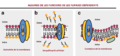 Funcions flipasa wiki 1.png 1,196 × 559; 187 KB
-
 Funcions flipasa wiki.png 1,196 × 559; 176 KB
-
 Funcions flipases.png 748 × 373; 108 KB
-
Functional-Loss-of-Bmsei-Causes-Thermosensitive-Epilepsy-in-Contractile-Mutant-Silkworm-Bombyx-mori-srep12308-s1.ogv 1 min 0 s, 851 × 480; 4.69 MB
-
-
-
-
-
-
-
-
-
-
-
-
-
-
-
 Human.gif 338 × 398; 12 KB
-
 Hypertonic Isotonic Hypotonic.png 2,218 × 700; 101 KB
-
-
-
Impact-of-Lipid-Composition-and-Receptor-Conformation-on-the-Spatio-temporal-Organization-of-μ-pcbi.1005240.s024.ogv 1 min 19 s, 1,014 × 948; 70.68 MB
-
-
-
-
-
-
-
-
In-vivo-single-molecule-imaging-identifies-altered-dynamics-of-calcium-channels-in-dystrophin-ncomms5974-s5.ogv 3.9 s, 1,024 × 512; 6.83 MB
-
-
In-vivo-single-molecule-imaging-identifies-altered-dynamics-of-calcium-channels-in-dystrophin-ncomms5974-s7.ogv 9.5 s, 768 × 1,024; 48.12 MB
-
-
-
Inhomogeneity-Based-Characterization-of-Distribution-Patterns-on-the-Plasma-Membrane-pcbi.1005095.s017.ogv 5.0 s, 1,450 × 1,743; 1.16 MB
-
Intrinsically-disordered-proteins-drive-membrane-curvature-ncomms8875-s2.ogv 4.0 s, 228 × 228; 80 KB
-
Intrinsically-disordered-proteins-drive-membrane-curvature-ncomms8875-s3.ogv 4.2 s, 294 × 147; 56 KB
-
Intrinsically-disordered-proteins-drive-membrane-curvature-ncomms8875-s4.ogv 4.2 s, 512 × 256; 1.04 MB
-
-
-
-
-
-
Key-Role-of-Local-Regulation-in-Chemosensing-Revealed-by-a-New-Molecular-Interaction-Based-Modeling-pcbi.0020082.sv001.ogv 1 min 48 s, 432 × 360; 1.96 MB
-
Key-Role-of-Local-Regulation-in-Chemosensing-Revealed-by-a-New-Molecular-Interaction-Based-Modeling-pcbi.0020082.sv002.ogv 3 min 17 s, 432 × 360; 1.88 MB
-
Key-Role-of-Local-Regulation-in-Chemosensing-Revealed-by-a-New-Molecular-Interaction-Based-Modeling-pcbi.0020082.sv003.ogv 2 min 39 s, 432 × 360; 1.44 MB
-
Key-Role-of-Local-Regulation-in-Chemosensing-Revealed-by-a-New-Molecular-Interaction-Based-Modeling-pcbi.0020082.sv004.ogv 6 min 26 s, 432 × 360; 3.62 MB
-
Key-Role-of-Local-Regulation-in-Chemosensing-Revealed-by-a-New-Molecular-Interaction-Based-Modeling-pcbi.0020082.sv005.ogv 8 min 42 s, 288 × 240; 3.93 MB
-
Key-Role-of-Local-Regulation-in-Chemosensing-Revealed-by-a-New-Molecular-Interaction-Based-Modeling-pcbi.0020082.sv006.ogv 4 min 17 s, 288 × 240; 3.36 MB
-
-
Lateral-Diffusion-on-Tubular-Membranes-Quantification-of-Measurements-Bias-pone.0025731.s005.ogv 33 s, 436 × 155; 2.36 MB
-
Lateral-Diffusion-on-Tubular-Membranes-Quantification-of-Measurements-Bias-pone.0025731.s006.ogv 8.1 s, 320 × 235; 373 KB
-
Lectin-Based-Food-Poisoning-A-New-Mechanism-of-Protein-Toxicity-pone.0000687.s001.ogv 5.2 s, 360 × 360; 61 KB
-
Lectin-Based-Food-Poisoning-A-New-Mechanism-of-Protein-Toxicity-pone.0000687.s002.ogv 5.2 s, 360 × 360; 131 KB
-
Lectin-Based-Food-Poisoning-A-New-Mechanism-of-Protein-Toxicity-pone.0000687.s003.ogv 5.2 s, 360 × 360; 370 KB
-
Lectin-Based-Food-Poisoning-A-New-Mechanism-of-Protein-Toxicity-pone.0000687.s004.ogv 5.2 s, 360 × 360; 298 KB
-
Lectin-Based-Food-Poisoning-A-New-Mechanism-of-Protein-Toxicity-pone.0000687.s005.ogv 5.2 s, 360 × 360; 123 KB
-
Lectin-Based-Food-Poisoning-A-New-Mechanism-of-Protein-Toxicity-pone.0000687.s006.ogv 5.2 s, 360 × 360; 52 KB


