Category:Pathology
Prijeđi na navigaciju
Prijeđi na pretraživanje

study and diagnosis of disease .jpg) | |||||
| Postavi datoteku | |||||
| Zvučni zapis izgovora | |||||
|---|---|---|---|---|---|
| Jest |
| ||||
| Dio klase |
| ||||
| Različito od | |||||
| |||||
Potkategorije
Ova kategorija ima sljedećih 50 potkategorija, od ukupno 50.
*
A
B
C
D
E
- Emperipolesis (4 F)
F
G
- General pathology (14 F)
H
I
L
M
- Media from PathoGenetics (1 F)
N
O
P
- Pathobiochemistry (2 F)
- Pathogenesis (6 F)
- Pathologie Kiel (2 F)
R
S
T
U
- UVS E building, Budapest (7 F)
V
Mediji u kategoriji »Pathology«
Prikazano je 70 datoteka u ovoj kategoriji, od njih ukupno 70.
-
 12-06-11-rechtsmedizin-berlin-08.jpg 3.216 × 2.136; 3,17 MB
-
 20071017010DR Dresden-Friedrichstadt Krankenhaus Pathologie.jpg 1.920 × 2.544; 6,18 MB
-
 Actinomycosis.pdf 1.239 × 1.752; 988 KB
-
 Anzar AYan.jpg 2.208 × 3.920; 785 KB
-
 Aster Labs.jpg 2.448 × 3.264; 2,11 MB
-
 Basal cell carcinoma histology image.jpg 2.464 × 3.188; 3,04 MB
-
 Chart showing development. Wellcome L0001433.jpg 1.688 × 1.229; 325 KB
-
 Covid-19 'Alert Level 4' pathology centre notices, Waikanae.jpg 2.985 × 1.884; 762 KB
-
 COVID-19-Antigen-with-Influenza-A-B-Rapid-Antigen-Combo.jpg 4.640 × 3.472; 4,17 MB
-
 A manual of pathology (IA cu31924000902878).pdf 662 × 1.056, 563 stranice; 14,73 MB
-

-
 Ear, nose and throat plaster casts, Sutton, England, 1890 Wellcome L0058781.jpg 2.832 × 3.994; 1,23 MB
-
 Effect on blood cells of removal of spleen Wellcome M0013298.jpg 5.139 × 2.152; 1,5 MB
-
 EIPH hemosiderin content in alveolar macrophages.jpg 2.137 × 239; 157 KB
-
 Faceted Search example.jpg 1.920 × 1.160; 458 KB
-
 FattTumor.png 600 × 428; 141 KB
-
 Fatty degeneration of certain tissues. Wellcome L0004773.jpg 1.868 × 1.052; 974 KB
-
 Figure 1 (11108271853).png 444 × 349; 327 KB
-
 FitoterapiaElementi.png 800 × 602; 236 KB
-
 Foundation of medicine-min.jpg 468 × 329; 56 KB
-
 Generalized perio -touched up.jpg 871 × 325; 29 KB
-
 Great Northern Central Hospital, Holloway Road, London; the Wellcome L0017726.jpg 1.504 × 1.234; 595 KB
-
 Great Northern Central Hospital, Holloway Road, London; the Wellcome V0028923.jpg 3.070 × 2.280; 2,19 MB
-
 GVDs Wiki Final Bar=20um.jpg 900 × 627; 945 KB
-
 Happy Halloween! (284183160).jpg 2.992 × 2.764; 3,81 MB
-
 Her2neu 3+staining.jpg 4.912 × 3.684; 3,68 MB
-
 Hospital Pathologist working at a computer.jpg 1.170 × 1.237; 818 KB
-
 Institut für Pathologie (Kiel).jpg 1.346 × 917; 388 KB
-
 Journal.pone.0026395.g001 cervical cytology brushes.png 1.345 × 1.788; 3,55 MB
-

-
 Laboratory improvement project in areas of Uganda.jpg 371 × 241; 31 KB
-
 Lipoma histology diagram.o.jpg 6.936 × 9.248; 12,73 MB
-
 LIPOMA spotter(General pathology ).jpg 4.624 × 3.468; 6,49 MB
-
 Margination of neutrophils in acute inflammation.png 724 × 540; 1.012 KB
-
 PAs cut at microscopy - extremely low mag.jpg 4.000 × 6.000; 9,07 MB
-
 Pathologisches Institut-Koenigsberg.jpg 3.000 × 2.260; 1,21 MB
-
 Pathology Collections.jpg 1.080 × 606; 30 KB
-
 Peec05.jpg 438 × 257; 10 KB
-
 Plant (fecal) material from colon biopsy (6032555128).jpg 1.630 × 1.086; 856 KB
-
 Post-mortem set, United Kingdom, 1860-1865 Wellcome L0058745.jpg 3.506 × 4.248; 2,42 MB
-
 Preparatul transilvanean, 1995, Aquarell auf Papier, 56 x 52 cm.jpg 944 × 1.024; 811 KB
-
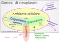
 PromotoriPsichiciNeoplasmi.png 600 × 571; 252 KB
-
 Quantitative parasitology 1.JPG 598 × 434; 26 KB
-
 ReferenzeEmatiche.jpg 400 × 112; 7 KB
-
 Rhinosporidiosis histology.jpg 837 × 1.097; 310 KB
-
 RHINOSPORIDIOSIS.jpg 2.479 × 2.958; 3,55 MB
-
 Rosen's Diagnosis of Breast Pathology by Needle Core Biopsy.jpg 2.953 × 3.939; 2,7 MB
-
 SCCOHT HE 20x.jpg 4.912 × 3.684; 3,66 MB
-
 Schwannoma Histopathology S100.jpg 4.912 × 3.684; 5,08 MB
-
 Südharz Klinikum Nordhausen - Hinterhof Pathologie.jpg 4.000 × 3.000; 2,74 MB
-
 Thagomizer (spike).jpg 2.100 × 2.685; 1,27 MB
-
 Thagomizer.tif 6.101 × 4.886; 28,45 MB
-
 Travelling case, T.J. Horder Wellcome M0019377.jpg 2.712 × 3.936; 2,05 MB
-
 Type 1 hypersensitivity v1.pdf 1.658 × 583; 41 KB
-
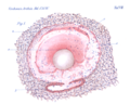 Uber das Vorkommen der D u er c k 1907 fig1.tiff 988 × 787; 1,11 MB
-
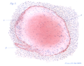 Uber das Vorkommen der D u er c k 1907 fig2.tiff 933 × 729; 1,12 MB
-
 Unstained peripheral blood smear closeup.jpg 5.184 × 3.456; 11,29 MB
-
 VariazLeucocitiInfezione.jpg 675 × 495; 220 KB
-
 Vibratome.jpg 1.536 × 2.048; 1,03 MB
-
 VT1200 S hero right 45 degrees.jpg 6.006 × 4.260; 1,38 MB
-
 Whole slide image of H&E stained breast tumour tissue.png 8.213 × 5.321; 34,44 MB
-
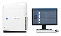 ZEISS Axio Scan.Z1 (8407041905).jpg 6.359 × 3.744; 1,4 MB
-
 Zeitschrift für Hygiene. Bd. LXXXVII Fig4.tiff 330 × 207; 69 KB
-
 Zeitschrift für Hygiene. Bd. LXXXVII Fig5.tiff 538 × 531; 459 KB
-
 Zeitschrift für Hygiene. Bd. LXXXVII Fig6.tiff 241 × 304; 88 KB
-
 Zeitschrift für Hygiene. Bd. LXXXVII Fig7.tiff 298 × 292; 76 KB
-
 Zeitschrift für Hygiene. Bd. LXXXVII Fig8.tiff 241 × 169; 47 KB
-
 Zeitschrift für Hygiene. Bd. LXXXVII Fig9.tiff 889 × 749; 1,07 MB
-
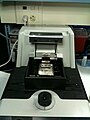
 ZEN browser for Virtual Microscopy (9318136341).jpg 4.235 × 3.123; 2,01 MB
-
 Дисплазія вушної раковини.jpg 3.024 × 4.032; 1,16 MB


_(14592593079).jpg)


.png)


.jpg)


.jpg)


.jpg)


_material_from_colon_biopsy_(6032555128).jpg)


.jpg)


.jpg)
.jpg)
