Category:Histopathology
Jump to navigation
Jump to search

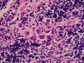
Histopathology is the microscopic examination of tissue in order to study the manifestations of disease.
microscopic examination of (live or dead) biological tissue samples in order to study and diagnose diseases   | |||||
| Upload media | |||||
| Instance of |
| ||||
|---|---|---|---|---|---|
| Subclass of | |||||
| |||||
- Place files relating to the diseased tissues of human beings in a suitable subcategory of "Category:Human histopathology".
- Place files relating to the diseased tissues of animals in a suitable subcategory of "Category:Veterinary histopathology".
- Place files relating to normal, disease-free tissue in a suitable subcategory of "Category:Animal histology", "Category:Human histology" or "Category:Plant histology".
Subcategories
This category has the following 31 subcategories, out of 31 total.
.
?
- Histopathology - unidentified (135 F)
A
- Atypia (1 F)
C
- Corpora amylacea (8 F)
G
- Gandy–Gamna nodules (3 F)
- Giant cells (4 F)
H
- Histopathology of cell death (1 F)
- Histopathology of lipofuscin (6 F)
I
M
N
O
P
R
- Russell bodies (8 F)
S
T
Pages in category "Histopathology"
This category contains only the following page.
Media in category "Histopathology"
The following 200 files are in this category, out of 293 total.
(previous page) (next page)-
 12882 2020 1959 Fig5 HTML (1).webp 685 × 513; 127 KB
-
 13071 2019 3727 Fig6.webp 1,590 × 1,851; 850 KB
-
 19-0448-F1.jpg 1,500 × 1,101; 201 KB
-
 Actinogranuloma (27484829990).jpg 1,024 × 768; 204 KB
-
 Actinogranuloma (27728825886).jpg 943 × 707; 106 KB
-
 Air bubble entrapment artifact.png 1,316 × 754; 2.12 MB
-
 Axillary Lymph Node Containing Gold Used To Treat Rheumatoid Arthritis (49230029317).jpg 1,761 × 1,196; 1.05 MB
-
 Axillary Lymph Node with Black (Tattoo?) Pigment (17509353414).jpg 2,048 × 1,536; 1,013 KB
-
 Barium- Pericolonic (49739606498).jpg 1,936 × 1,936; 1.19 MB
-
 Barium-Colonic Perforation (49750169036).jpg 1,936 × 1,936; 1.17 MB
-
 Bone Cells (5828753702).jpg 1,325 × 1,013; 1.38 MB
-
 Breast Cancer and Alcohol Consumption (5881108114).jpg 1,874 × 1,800; 2.55 MB
-
 Breast Cancer; Nanotubes; Antibodies (5880985350).jpg 839 × 360; 202 KB
-
 Brisk Extravascular Hemolytic Anemia (24467236849).jpg 1,428 × 1,426; 1 MB
-
 CFHR5N C3 Immuno EM.tif 300 × 303; 290 KB
-
-
 CIDP Histopathology Teased fibre.jpg 1,040 × 772; 83 KB
-
 Citolisis por Doderlein (9138808173).jpg 640 × 480; 199 KB
-
 Citolisis por Doderlein (9141037502).jpg 640 × 480; 232 KB
-

-
 Clogged Pore on Nose Seen Through 100x Magnification Microscope.jpg 1,578 × 1,574; 391 KB
-
 Cluster of epithelium shedding from gingiva around dental implant.jpg 640 × 480; 210 KB
-
 Cryptococcus in cerebrospinal fluid, India ink prep. (49623531757).jpg 1,659 × 1,938; 1.26 MB
-
 Cutaneous Keratocyst (50267985312).jpg 2,107 × 2,124; 1.99 MB
-

-

-

-

-
 Drug-induced liver injury (DILI).png 450 × 680; 792 KB
-
 Echinococcus granulosus hooklet x400 mag (1) (7686892792).jpg 1,024 × 768; 138 KB
-
 Echinococcus granulosus hooklet x400 mag (2) (7686893156).jpg 1,024 × 768; 145 KB
-

-

-
 Epithelium cell (33390275448).jpg 2,560 × 1,920; 2.01 MB
-
 Eustachian tube.tif 638 × 438; 570 KB
-

-
-
 Extendido atrófico (9138805703).jpg 640 × 480; 146 KB
-
 Extendido atrófico (9138805727).jpg 640 × 480; 219 KB
-
 Extendido atrófico (9141035068).jpg 640 × 480; 121 KB
-
 Extendido atrófico (9438100071).jpg 1,280 × 960; 442 KB
-
 Extendido atrófico (9438100077).jpg 1,280 × 960; 544 KB
-
 Extendido atrófico (9438100111).jpg 1,280 × 960; 570 KB
-
 Extendido de células superficiales e intermedias (9138691607).jpg 1,280 × 960; 527 KB
-
 Extendido de células superficiales e intermedias (9140919248).jpg 1,280 × 960; 501 KB
-
 Extendido luteínico (9399641333).jpg 1,280 × 960; 554 KB
-
 Extendido luteínico (9402403132).jpg 1,280 × 960; 571 KB
-
 F0841839 (27728823426).jpg 1,024 × 768; 229 KB
-
 F11631279 (27728816806).jpg 904 × 654; 193 KB
-
 F18057583 (27763115545).jpg 1,003 × 752; 266 KB
-
 F3361903 (27728820266).jpg 1,024 × 768; 237 KB
-
 Fig2--catenin-is-aberrant-expressed-in-cytoplasm-of-ABs-by-SP-20.jpg 359 × 269; 41 KB
-
 Figure 2 (6816966284).png 560 × 860; 1.1 MB
-
 Figure 2 (6900617586).png 913 × 524; 462 KB
-
 Figure 2 (7040513261).png 654 × 653; 830 KB
-
 Figure 2 (7046713495).png 946 × 731; 1.68 MB
-
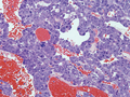 Figure 2 (7192020508).png 910 × 686; 1.62 MB
-
 Figure 2 (7282700658).png 788 × 615; 1.06 MB
-
 Figure 2 (7497587350).png 761 × 570; 1.12 MB
-
 Figure 2 (7978640678).png 864 × 361; 385 KB
-

-
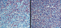 Figure 3 (6894418468).png 911 × 435; 1.07 MB
-
 Figure 3 (6948823394).png 606 × 523; 248 KB
-
 Figure 3 (7001446681).png 926 × 237; 281 KB
-
 Figure 3 (7797994594).png 873 × 651; 1.26 MB
-
 Figure 3B (6890588460).png 786 × 599; 563 KB
-
 Figure 3B (7769083744).png 865 × 612; 689 KB
-
 Figure 3B (7890285008).png 771 × 823; 629 KB
-
 Figure 4 (6873190814).png 676 × 859; 96 KB
-
 Figure 4 (6926214470).png 796 × 528; 894 KB
-
 Figure 4 (7036682837).png 848 × 823; 1.38 MB
-
 Figure 4 (7104078935).png 897 × 561; 411 KB
-
 Figure 4 (7346440130).png 774 × 779; 920 KB
-
 Figure 4 (7700731370).png 591 × 911; 1.28 MB
-
 Figure 4 (7736269166).png 625 × 866; 1.13 MB
-
 Figure 4 (7749859150).png 882 × 663; 1.32 MB
-
 Figure 4 (7784239892).png 848 × 631; 1.45 MB
-
 Figure 4 (7834330442).png 561 × 891; 1.12 MB
-
 Figure 4 (7993575877).png 671 × 777; 1.03 MB
-
 Figure 4 (8072006620).png 777 × 608; 786 KB
-
 Figure 4A (8123172312).png 727 × 704; 790 KB
-
 Figure 4B (7652696002).png 848 × 625; 1.12 MB
-
 Figure 4C (7890284262).png 811 × 872; 1.16 MB
-
 Figure 4D (7363493792).png 883 × 664; 1.25 MB
-
 Figure 5 (6798816584).png 869 × 548; 495 KB
-
 Figure 5 (6852655590).png 931 × 580; 484 KB
-
 Figure 5 (6882049750).png 652 × 882; 1.05 MB
-
 Figure 5 (6994454003).png 903 × 377; 202 KB
-
 Figure 5 (7416722982).png 679 × 679; 640 KB
-
 Figure 5 (8117099321).png 623 × 530; 897 KB
-
 Figure 6 (7245535388).png 608 × 646; 795 KB
-

-
 Freezing (ice crystal) artifact (7493541268).jpg 2,560 × 1,920; 2.62 MB
-

-

-

-
 Hales colloidal iron staining.jpg 2,040 × 1,536; 1.16 MB
-
 HEPARGF.jpg 1,936 × 1,456; 564 KB
-
 HepaRGUndiff.jpg 1,936 × 1,456; 514 KB
-

-

-
 Hongos (9138770107).jpg 640 × 480; 197 KB
-
 Hongos (9138770341).jpg 640 × 480; 154 KB
-
 Hongos (9138770877).jpg 640 × 480; 132 KB
-
 Hongos (9138771273).jpg 640 × 480; 147 KB
-
 Hongos (9140998948).jpg 640 × 480; 186 KB
-
 Hongos (9140999724).jpg 640 × 480; 152 KB
-
 Hongos (9141000614).jpg 640 × 480; 166 KB
-
 Hongos (9316824137).jpg 1,280 × 960; 430 KB
-
 Hongos (9316824285).jpg 1,280 × 960; 500 KB
-
 Hongos (9319612646).jpg 1,280 × 960; 459 KB
-
 Hongos (9319612656).jpg 1,280 × 960; 455 KB
-
 Hongos (9319612672).jpg 1,280 × 960; 440 KB
-
 Hongos (9319612786).jpg 1,280 × 960; 411 KB
-
 Hongos (9388317119).jpg 1,280 × 960; 344 KB
-
 Hongos (9388317143).jpg 1,280 × 960; 346 KB
-
 Hongos (9388317153).jpg 1,280 × 960; 335 KB
-
 Hongos (9391090480).jpg 1,280 × 960; 358 KB
-
 HPV-Infected Squamous Cell (Koilocyte) of the Cervix (44001403165).jpg 2,480 × 1,967; 744 KB
-
 Hyaluronic Acid - Breast (49724641207).jpg 960 × 500; 245 KB
-
 Hyaluronic Acid - Lip (49416531962).jpg 1,280 × 720; 196 KB
-
 Hyaluronic acid - Skin (50829198997).jpg 3,384 × 2,708; 1,020 KB
-

-

-
-

-
-

-

-
-

-

-
 Inclusion station.jpg 2,304 × 1,536; 530 KB
-
 Incomplete fixation.jpg 1,969 × 2,689; 1.11 MB
-

-

-

-
 Intramuscular Lipoma (38578598922).jpg 1,814 × 1,673; 994 KB
-

-
-
 Invasive Lobular Carcinoma of the Breast (33768522968).jpg 1,405 × 705; 156 KB
-
-

-
 Lamella bone H&E and under polarised light.gif 1,000 × 750; 684 KB
-
 Large Solitary Fibrous Tumor in the Retroperitoneum (8088106847).png 810 × 609; 1.25 MB
-
 Large Solitary Fibrous Tumor in the Retroperitoneum (8095041151).png 812 × 607; 862 KB
-

-
 Leukoplakia histology.jpg 448 × 380; 49 KB
-

-
 Marble spleen diasease (27728945806).jpg 1,024 × 768; 353 KB
-
 Megakaryocyte emboli.jpg 1,280 × 960; 261 KB
-
 Mesial sclerosis Neurfilament.jpg 2,080 × 1,542; 619 KB
-
 Metallosis (50618218978).jpg 454 × 680; 178 KB
-
 Microangiopathic Hemolytic Anemia (49129314097).jpg 2,057 × 2,250; 795 KB
-
 Mild-polyneuritis-rhesus-macaque--Macaca-mulatta.png 1,079 × 861; 985 KB
-

-
-
 Monochorionic Diamniotic Twins, Intervening Membrane.jpg 1,005 × 1,786; 801 KB
-
 Monosodium Urate Crystals in Elbow Joint Fluid (43911233991).jpg 2,052 × 2,172; 1.39 MB
-

-

-

-
-
 Mucoid impaction of bronchi associated with Bipolaris sp. (5136249993).jpg 1,280 × 960; 601 KB
-

-
 Nenadorove zmeny.svg 720 × 405; 34 KB
-
 Nochnitsa geminidens holotype right side.png 3,750 × 1,556; 6.02 MB
-
 Non-neoplastic changes.svg 720 × 405; 34 KB
-
 NonneoplasidrawDH.JPG 653 × 257; 22 KB
-

-

-
 P53-immunostaining-in-malignant-breast-epithelial-cells.jpg 600 × 490; 261 KB
-

-
 Paper in Colonic Lumen - Pica (50073036767).jpg 920 × 703; 297 KB
-
 Paraganglioma of Prostatic Origin (7933276588).png 829 × 432; 941 KB
-
 Parasite170122 Figs 20-25 Cavisoma magnum (Acanthocephala).png 5,100 × 6,600; 11.83 MB
-
 Pared del corazón.jpg 1,392 × 1,040; 327 KB
-
 Pathological Rupture of the Spleen in Uncomplicated Myeloma (7258460326).png 888 × 665; 1.55 MB
-
 Pathology of Gastrointestinal Stromal Tumors (8064498505).png 801 × 690; 1.26 MB
-
 Pathology of Gastrointestinal Stromal Tumors (8095041835).png 700 × 599; 1.04 MB
-
 Pathology of Gastrointestinal Stromal Tumors (8123155091).png 612 × 520; 691 KB
-

-
 PAX5 immunohistochemistry in relapsed classical Hodgkin's lymphoma.jpg 1,600 × 1,200; 1,007 KB
-

-
 Peritoneal inclusion cystx100.jpg 1,392 × 1,040; 1,012 KB
-
 Peritoneal inclusion cystx20.jpg 1,392 × 1,040; 865 KB
-

-
 PET Hyaluronic Acid Tissue.png 273 × 166; 129 KB
-
 Phaeohyphomycosis of the Finger (50436074978).jpg 2,505 × 1,694; 5.82 MB
-
 Photomicrograph of colorectal medullary carcinoma x20a.jpg 2,560 × 1,920; 3.21 MB
-
 Photomicrograph of colorectal medullary carcinoma x20d.jpg 2,560 × 1,920; 3.38 MB
-
 Photomicrograph of medullary carcinoma at anorectum x20a.jpg 2,560 × 1,920; 2.71 MB
-
 Photomicrograph of medullary carcinoma at anorectum x4a.jpg 2,560 × 1,920; 3.11 MB
-
 Pierre Masson(3).jpg 600 × 949; 172 KB
-

-
 Preparat cytologiczny wymazu z szyjki macicy.jpg 434 × 346; 28 KB
-
 Proteasome Inhibition - Thinking Outside the Box (7126489699).png 903 × 520; 269 KB
-
 Pseudoactinomycotic Radiate Granule (PAMRAG) (46228772372).jpg 1,827 × 1,347; 1.04 MB
-
 Pseudoactinomycotic Radiate Granules (PAMRAG) (50276883556).jpg 2,480 × 1,490; 1.71 MB
-

.webp)


.jpg)
.jpg)

.jpg)
_Pigment_(17509353414).jpg)
.jpg)
.jpg)
.jpg)
.jpg)
.jpg)
.jpg)


.jpg)
.jpg)
.png)


.jpg)
.jpg)
.png)
.png)
.png)
.png)
.png)
_(7686892792).jpg)
_(7686893156).jpg)
.png)
.jpg)
.png)
.png)
.jpg)
.jpg)
.jpg)
.jpg)
.jpg)
.jpg)
.jpg)
.jpg)
.jpg)
.jpg)
.jpg)
.jpg)
.jpg)
.jpg)

.png)
.png)
.png)
.png)
.png)
.png)
.png)
.png)
.png)
.png)
.png)
.png)
.png)
.png)
.png)
.png)
.png)
.png)
.png)
.png)
.png)
.png)
.png)
.png)
.png)
.png)
.png)
.png)
.png)
.png)
.png)
.png)
.png)
.png)
.png)
.png)
.png)
.png)
.png)
_artifact_(7493541268).jpg)
.png)
.png)


_Caused_by_Unbalanced_Diet_(8048667027).png)
_Caused_by_Unbalanced_Diets_(8072012183).png)
.jpg)
.jpg)
.jpg)
.jpg)
.jpg)
.jpg)
.jpg)
.jpg)
.jpg)
.jpg)
.jpg)
.jpg)
.jpg)
.jpg)
.jpg)
.jpg)
.jpg)
_of_the_Cervix_(44001403165).jpg)
.jpg)
.jpg)
.jpg)
.png)
.png)
.png)
.png)
.png)
.png)
.png)
.png)


.png)
.png)
.jpg)
.png)
.png)
.jpg)
.png)
.png)

.png)
.png)
.png)

.png)
.jpg)


.jpg)
.jpg)

.png)
.png)

.jpg)
.png)
.png)
_(7181752555).png)
_(8078164162).png)
.jpg)
.png)


.png)
.png)

.png)
.jpg)
.png)
.png)

.png)
.png)
.png)
.png)
.png)

.png)


.png)

.jpg)


.jpg)
.png)

.png)
_(46228772372).jpg)
_(50276883556).jpg)
.png)
.png)
.png)
.png)
.png)
.png)
.png)