File:Giardia 1.jpg
Jump to navigation
Jump to search

No higher resolution available.
Giardia_1.jpg (700 × 519 pixels, file size: 25 KB, MIME type: image/jpeg)
Captions
Captions
Add a one-line explanation of what this file represents
Summary
[edit]
| Description |
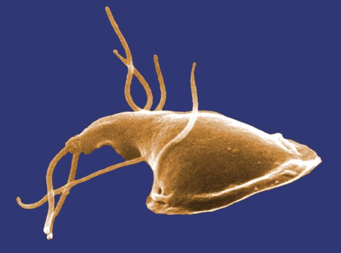
English: ID#: 11649
Description: This digitally-colorized scanning electron micrograph (SEM) depicted the dorsal (upper) surface of a Giardia protozoan that had been isolated from a rat’s intestine. Some of the identifying morphologic characteristics include pairs of thread-like flagella that facilitate motility, and a ventolateral flange that appears as a “ruffle” around the anterior portion of the organism. Pairs of flagella seen here include an anterior, posterior-lateral, and caudal pairs. The protozoan Giardia causes the diarrheal disease called giardiasis. Giardia species exist as free-swimming (by means of flagella) trophozoites, and as egg-shaped cysts. It is the cystic stage, which facilitates the survival of these organisms under harsh environmental conditions. The cyst is considered the infective form, and disease is often transmitted by drinking contaminated water. As depicted in these SEMs, in the intestine, cysts are stimulated to liberate trophozoites. Cysts can be shed in fecal material, and can, thereafter, remain viable for several months in appropriate environmental conditions. Cysts can also be transferred directly from person-to-person, as a result of poor hygiene. Dr. Stan Erlandsen; Dr. Dennis Feely |
| Date | |
| Source | http://phil.cdc.gov/PHIL_Images/11649/11649_lores.jpg |
| Author | Dr. Stan Erlandsen; Dr. Dennis Feely,Center for Disease Control |
Licensing
[edit]
This image is a work of the Centers for Disease Control and Prevention, part of the United States Department of Health and Human Services, taken or made as part of an employee's official duties. As a work of the U.S. federal government, the image is in the public domain.
|
File history
Click on a date/time to view the file as it appeared at that time.
| Date/Time | Thumbnail | Dimensions | User | Comment | |
|---|---|---|---|---|---|
| current | 06:45, 24 March 2010 | | 700 × 519 (25 KB) | 7mike5000 (talk | contribs) | {{Information |Description={{en|1=giardia}} |Source=http://phil.cdc.gov/PHIL_Images/11649/11649_lores.jpg |Author=Janice Harney Carr. Center for Disease Control |Date=2006 |Permission= |other_versions= }} Category:Insects |
You cannot overwrite this file.
File usage on Commons
There are no pages that use this file.
File usage on other wikis
The following other wikis use this file:
- Usage on bs.wikipedia.org
- Usage on ca.wikipedia.org
- Usage on en.wikipedia.org
- Usage on en.wikiversity.org
- User:Jtwsaddress42/Projects/Project 1
- User:Jtwsaddress42/Projects/Project 1/Parts
- User:Jtwsaddress42/Projects/Project 1/Parts/Part 4
- User:Jtwsaddress42/Projects/Project 1/Chapters/Chapter 11
- User:Jtwsaddress42/Projects/Project 1/Sections/Chapter 11/Phase I - The Rise of Neomurans and the Modern Aerobic Atmosphere (950-600 mya)
- User:Jtwsaddress42/Clade
- User:Jtwsaddress42/Clade/Neomura to Podiata
- Usage on es.wikipedia.org
- Usage on tr.wikipedia.org
- Usage on uk.wikipedia.org
- Usage on vi.wikipedia.org