File:Мозочок.jpg
Jump to navigation
Jump to search

Size of this preview: 800 × 600 pixels. Other resolutions: 320 × 240 pixels | 640 × 480 pixels | 1,024 × 768 pixels | 1,280 × 960 pixels | 2,560 × 1,920 pixels | 3,000 × 2,250 pixels.
Original file (3,000 × 2,250 pixels, file size: 7.13 MB, MIME type: image/jpeg)
Captions
Captions
Add a one-line explanation of what this file represents
Summary
[edit]
| Description |
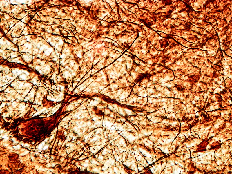
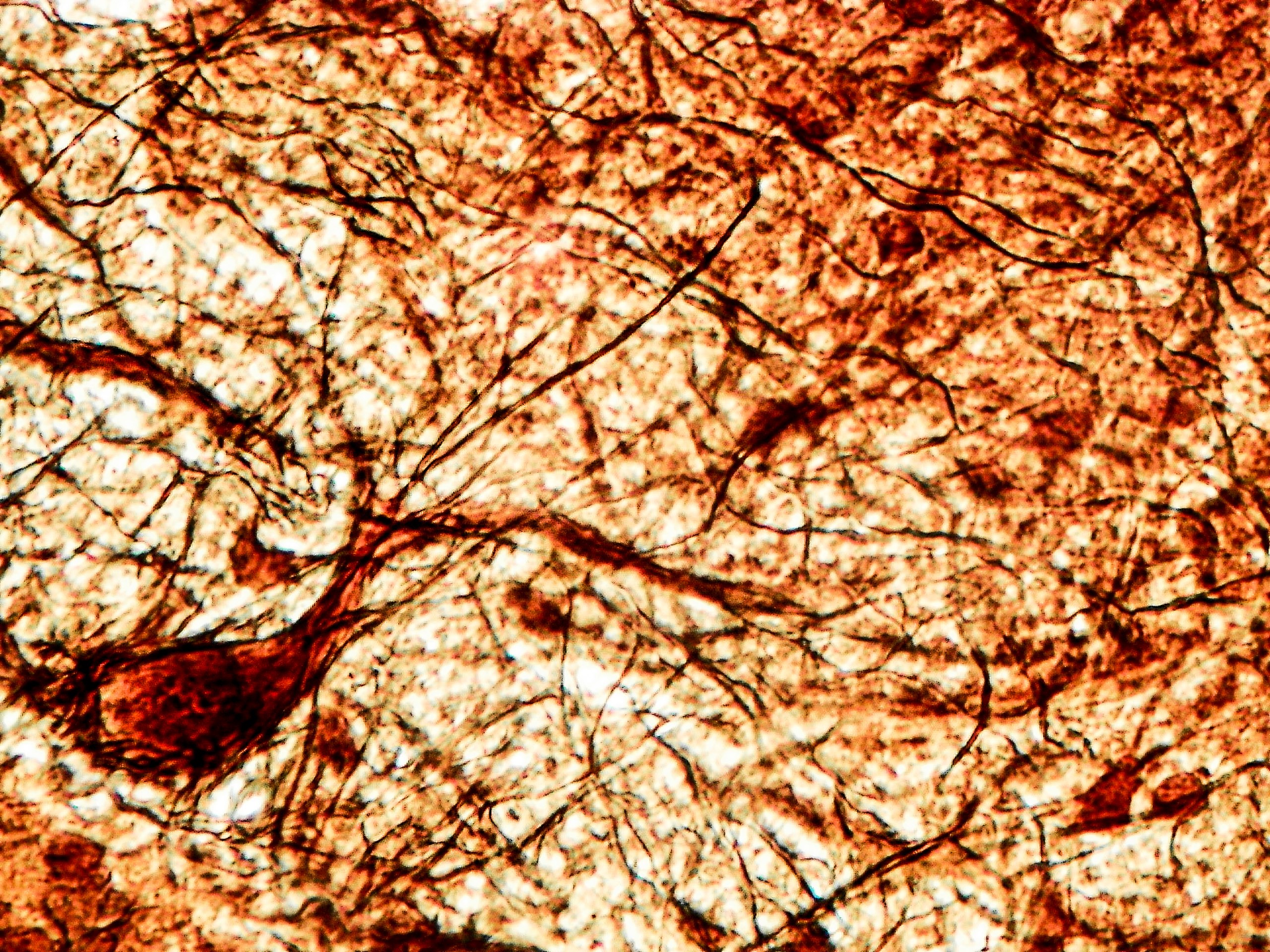
Українська: Кора мозочка.
Фарбування: імпрегнація азотно-кислим сріблом. Збільшення: 400. Мікроскоп: MICROmed (XS-5520). Кора мозочка включає три шари: 1. Молекулярний шар – найбільш поверхневий; утворений дендритними кронами клітин Пуркіньє, аксонами зернистих нейронів, тілами та відростками кошикових та зірчастих нейронів, клітинами-канделябрами; у нього також заходять дендрити клітин Гольджі; 2.Шар клітин Пуркіньє не суцільний - він утворений перикаріонами грушеподібних нейронів, які розташовані на певній відстані один від одного; 3.Зерничтий шар – безпосередньо прилеглий до білої речовини; основним видом клітин являються клітини-зерна, перикаріони яких можуть стикатися, формуючи окремі групи-кластери. Вільний простір між зернистими клітинами заповнений клітинами Гольджі, Лугаро, а також щіточковими нейронами.English: Cerebellar cortex. Dyeing: impregnation with nitric acid silver. Magnification: 400. Microscope: MICROmed (XS-5520). The cerebellar cortex includes three layers: 1. Molecular layer - the most superficial; formed by dendritic crowns of Purkinje cells, axons of granular neurons, bodies and processes of basket and stellate neurons, candelabra cells; it also includes dendrites of Golgi cells; 2. The layer of Purkinje cells is not continuous - it is formed by perikaryons of pear-shaped neurons, which are located at a certain distance from each other; 3. Granular layer - directly adjacent to the white matter; the main type of cells are grain cells, the perikaryons of which can collide, forming separate groups-clusters. The free space between granular cells is filled with Golgi, Lugaro cells, and also brush neurons. |
| Date | |
| Source | Own work |
| Author | Орися Поліщук |
Licensing
[edit]
I, the copyright holder of this work, hereby publish it under the following license:
This file is licensed under the Creative Commons Attribution 4.0 International license.
- You are free:
- to share – to copy, distribute and transmit the work
- to remix – to adapt the work
- Under the following conditions:
- attribution – You must give appropriate credit, provide a link to the license, and indicate if changes were made. You may do so in any reasonable manner, but not in any way that suggests the licensor endorses you or your use.
| This image was uploaded as part of Science Photo Competition 2020 in Ukraine. |

|
This media file is uncategorized.
Please help improve this media file by adding it to one or more categories, so it may be associated with related media files (how?), and so that it can be more easily found.
Please notify the uploader with {{subst:Please link images|File:Мозочок.jpg}} ~~~~ |
File history
Click on a date/time to view the file as it appeared at that time.
| Date/Time | Thumbnail | Dimensions | User | Comment | |
|---|---|---|---|---|---|
| current | 17:30, 29 November 2020 | | 3,000 × 2,250 (7.13 MB) | Орися Поліщук (talk | contribs) | Uploaded own work with UploadWizard |
You cannot overwrite this file.
File usage on Commons
There are no pages that use this file.