File:Плазмолиз красного лука.jpg
Original file (1,920 × 1,080 pixels, file size: 1.97 MB, MIME type: image/jpeg)
Captions
Captions
Summary
[edit]
| Description |
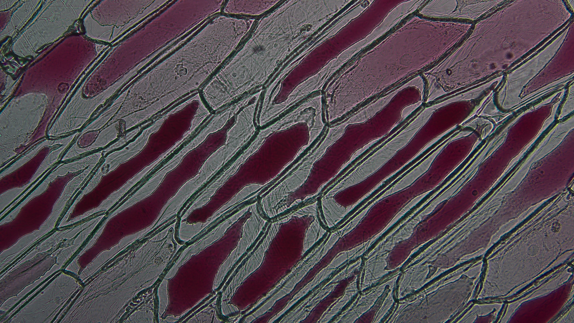
Русский: Плазмолиз красного лука (Plasmolysis of red onion)
Плазмолиз – это процесс отделения протопласта от клеточной стенки в гипертоническом растворе. Для демонстрации плазмолиза, нам потребуется препарат из кожицы красного лука, а также гипертонический и гипотонический раствор. Этапы работы: 1. Сначала надо изготовить гипертонический и гипотонический раствор. 2. Аккуратно снять кожицу лука с той стороны чешуи, где много пигмента. 3. Протереть стекла салфеткой, на предметное стекло добавить каплю воды и в ней расправить кожицу лука, накрыть покровным стеклом. Излишек воды оттянуть фильтровальной бумагой. 4. Поместить препарат на предметный столик под микроскоп и начать исследование с малого увеличения. Антоцианы – это водорастворимый пигмент, поэтому можно заметить, что при удалении излишка воды с препарата, вместе с водой вывелось часть пигмента из клетки, и салфетка окрасилась в малиновый цвет. Причина этому – осмос. 5. С одной стороны пипеткой следует добавить на препарат гипертонический раствор, а с другой стороны подложить фильтровальную бумагу. Вода активно будет перемещаться в сторону салфетки. В этот момент происходит: 1. Под действием высокого осмотического давления вода из клеток диффундирует в межклеточное пространство. 2. Вследствие выхода воды из клетки объем клеточного сока сокращается, тургор уменьшается. 3. Объем клеточной вакуоли уменьшается. 4. Наблюдаем процесс отслоения протопласта от клеточной стенки – это и есть плазмолиз. Микрофотография выполнена с использованием светового микроскопа Primo Star, Carl Zeiss в Центре выявления и поддержки одаренных детей "Гагарин" (Оренбург).English: Plasmolysis of red onion
Plasmolysis is the process of separating the protoplast from the cell wall in a hypertonic solution. To demonstrate plasmolysis, we will need a preparation from the skin of red onion, as well as a hypertonic and hypotonic solution. Stages of work: 1. First you need to make a hypertonic and hypotonic solution. 2. Carefully remove the onion skin from the side of the scales where there is a lot of pigment. 3. Wipe the glass with a napkin, add a drop of water to the slide and spread the onion skin in it, cover with a cover glass. Remove excess water with filter paper. 4. Place the drug on the slide table under the microscope and start the study with a small magnification. Anthocyanins are a water–soluble pigment, so you can notice that when removing excess water from the drug, part of the pigment was removed from the cell along with the water, and the napkin turned crimson. The reason for this is osmosis. 5. On the one hand, a hypertonic drug should be added to the drug with a pipette At this moment , the: 1. Under the influence of high osmotic pressure, water from the cells diffuses into the intercellular space. 2. Due to the release of water from the cell, the volume of cell juice is reduced, turgor decreases. 3. The volume of the cellular vacuole decreases. 4. We observe the process of detachment of the protoplast from the cell wall – this is plasmolysis. The micrograph was performed using a Primo Star, Carl Zeiss light microscope at the Gagarin Center for the Identification and Support of Gifted Children (Orenburg).English: Plasmolysis of red onion |
| Date | |
| Source | Own work |
| Author | Suchkova |
Licensing
[edit]
- You are free:
- to share – to copy, distribute and transmit the work
- to remix – to adapt the work
- Under the following conditions:
- attribution – You must give appropriate credit, provide a link to the license, and indicate if changes were made. You may do so in any reasonable manner, but not in any way that suggests the licensor endorses you or your use.
This image was uploaded as part of Russian Science Photo Competition 2023.
|
File history
Click on a date/time to view the file as it appeared at that time.
| Date/Time | Thumbnail | Dimensions | User | Comment | |
|---|---|---|---|---|---|
| current | 13:24, 31 May 2023 | | 1,920 × 1,080 (1.97 MB) | Suchkova (talk | contribs) | Uploaded own work with UploadWizard |
You cannot overwrite this file.
File usage on Commons
There are no pages that use this file.