File:2012 Sombke et al f04.png
Jump to navigation
Jump to search

Size of this preview: 452 × 599 pixels. Other resolutions: 181 × 240 pixels | 362 × 480 pixels | 579 × 768 pixels | 772 × 1,024 pixels | 2,005 × 2,658 pixels.
Original file (2,005 × 2,658 pixels, file size: 7.16 MB, MIME type: image/png)
Captions
Captions
Add a one-line explanation of what this file represents
Summary[edit]
| Description |
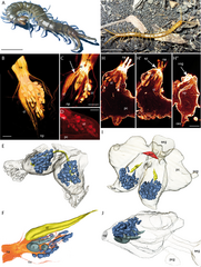
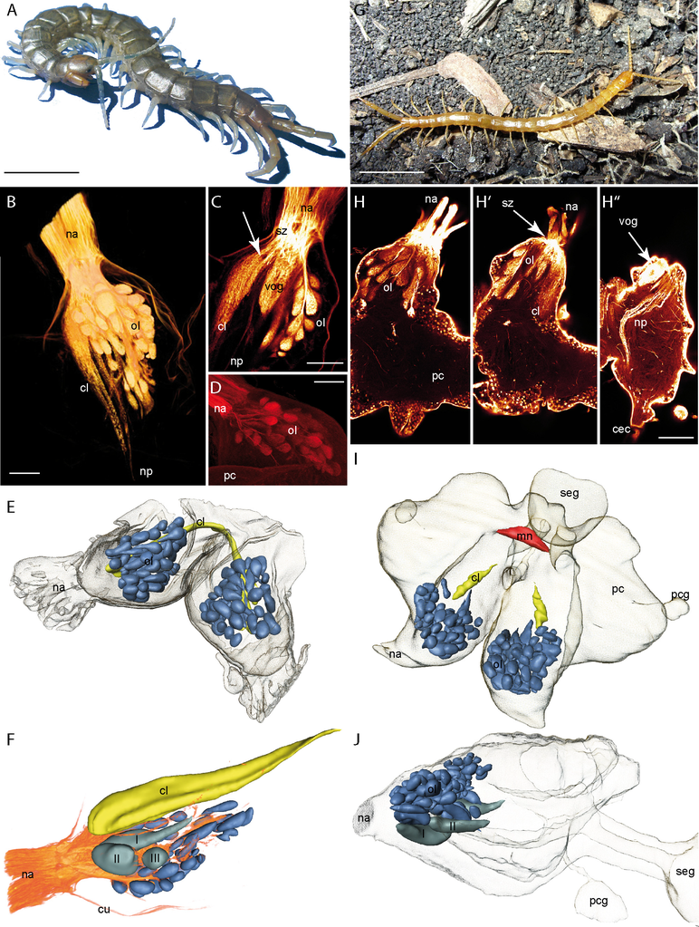
English: Figure 4 Scolopendromorpha. A Scolopendra oraniensis. B Neurobiotin backfill of the antennal nerve in S. oraniensis showing the olfactory lobe, the corpus lamellosum, and neurite projections (horizontal maximal projection, cLSM scan). C Single optical horizontal section of a Lucifer yellow backfill in S. oraniensis (cLSM scan). Antennal neurites cross each other in a sorting zone and project into different neuropils. The arrow marks the structural composition of the corpus lamellosum in which single lamellae are weakly noticeable. The large ventral OG is visible in this section. Single olfactory glomeruli in the olfactory lobe are arranged like in a grape. D Neurobiotin backfill of the antennal nerve of S. oraniensis. Only a subpopulation of the antennal neurites and olfactory glomeruli is labeled (horizontal maximal projection, cLSM scan). E 3D reconstruction of the brain of Scolopendra subspinipes (dorsal protocerebrum is not shown) with deutocerebral neuropils. Blue = olfactory glomeruli, yellow = corpus lamellosum. F 3D reconstruction of deutocerebral neuropils of Scolopendra oraniensis combined with volume rendering of the antennal backfill in B. Three enlarged ventral glomeruli (I, II, III) are present. G Cryptops hortensis. H Single horizontal optical sections (cLSM) of a neurobiotin backfill of the right antennal nerve in C. hortensis from dorsal to ventral. Antennal nerve bundles and innervation of single olfactory glomeruli. H’ Sorting zone (arrow) of antennal neurites and corpus lamellosum. H’’ Larger ventral olfactory glomerulus (arrow) and neurite projections. I 3D reconstruction of the brain of C. hortensis with deutocerebral neuropils and midline neuropil. Contralateral connection of the CL is not shown. Blue = olfactory glomeruli, yellow = corpus lamellosum, red = midline neuropil. J Lateral view of the 3D reconstruction in I. Two enlarged ventral glomeruli (I, II) are present. Abbreviations: cec circumesophageal connectives, cl corpus lamellosum, mn midline neuropil, na nervus antennalis, np neurite projections, ol olfactory lobe, pc protocerebrum, pcg protocerebral gland, seg subesophageal ganglion, sz sorting zone, vog ventral olfactory glomerulus. Scalebars: A and F = 10 mm, B-D, G = 100 μm. |
| Date | |
| Source | Sombke, Andy; Lipke, Elisabeth; Kenning, Matthes; Müller, Carsten HG; Hansson, Bill S.; Harzsch, Steffen (2012-01-03). Comparative analysis of deutocerebral neuropils in Chilopoda (Myriapoda): implications for the evolution of the arthropod olfactory system and support for the Mandibulata concept. BMC Neuroscience 13 (1): 1. doi:10.1186/1471-2202-13-1 |
| Author | Andy Sombke, Elisabeth Lipke, Matthes Kenning, Carsten HG Müller, Bill S Hansson, Steffen Harzsch |
Licensing[edit]
This file is licensed under the Creative Commons Attribution 2.0 Generic license.
- You are free:
- to share – to copy, distribute and transmit the work
- to remix – to adapt the work
- Under the following conditions:
- attribution – You must give appropriate credit, provide a link to the license, and indicate if changes were made. You may do so in any reasonable manner, but not in any way that suggests the licensor endorses you or your use.
File history
Click on a date/time to view the file as it appeared at that time.
| Date/Time | Thumbnail | Dimensions | User | Comment | |
|---|---|---|---|---|---|
| current | 01:09, 2 September 2022 | | 2,005 × 2,658 (7.16 MB) | Junnn11 (talk | contribs) | Uploaded a work by Andy Sombke, Elisabeth Lipke, Matthes Kenning, Carsten HG Müller, Bill S Hansson, Steffen Harzsch from Sombke, Andy; Lipke, Elisabeth; Kenning, Matthes; Müller, Carsten HG; Hansson, Bill S.; Harzsch, Steffen (2012-01-03). [https://www.researchgate.net/publication/51980717 Comparative analysis of deutocerebral neuropils in Chilopoda (Myriapoda): implications for the evolution of the arthropod olfactory system and support for the Mandibulata concept]. BMC Neuroscience 13 (1):... |
You cannot overwrite this file.
File usage on Commons
There are no pages that use this file.