File:3D Dual Color Super Resolution Microscopy Cremer 2010.png

原始檔案 (3,486 × 1,280 像素,檔案大小:3 MB,MIME 類型:image/png)
說明
說明
摘要
[編輯]| 描述 |
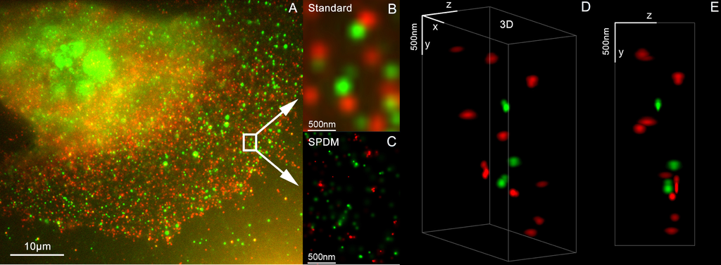
English: 3D Dual Color Super Resolution Microscopy by combining localization microscopy SPDM with spatially modulated illumination SMI / Optical nanoscopy / Christoph Cremer emeritus at Heidelberg university [1], [2]
3D reconstruction of dual color acquistion of Her2/neu and Her 3. A) conventional wide-field fluorescence image of an AG11132 mammary epithelial cells. Her3 is labelled with Alexa488 (green) and Her2/neu with Alexa568 (red). B) magnified image of a small area of A. C) localization microscopy SPDM of the same region of interest as in B. D) and E) show a 3D reconstruction of the protein clusters using the combination of SPDM and SMI microscopy (LIMON technology). Scale bars in D) and E) are 500 nm in each direction. By combining SPDMphymod with SMI (both invented in Christoph Cremer´s lab in 1996) a 3D dual colour reconstruction of the spatial arrangements of Her2/neu and Her3 clusters was achieved. The positions in all three directions of the protein clusters could be determined with an accuracy of about 25 nm. Her2/neu and Her3 are tyrosine kinase receptors and their overexpression is related to certain types breast cancer. The paper analyses the Her2 distribution pattern in breast cancer cells and counts the clusters (20 637 clusters with a mean diameter of 67 nm) with a specifically developed clusters finding algorithm for super resolution microscopy. This methods could help for the exploration of the resistance mechanism in Trastuzumab/Herceptin treatment. Publication: Rainer Kaufmann, Patrick Müller, Georg Hildenbrand, Michael Hausmann & Christoph Cremer [3], [4] : Analysis of Her2/neu membrane protein clusters in different types of breast cancer cells using localization microscopy, Journal of Microscopy 2010, doi: 10.1111/j.1365-2818.2010.03436.x |
| 日期 | 051611 |
| 來源 | 自己的作品 |
| 作者 | Andy Nestl |
Gallery
[編輯]- Super Resolution Microscopy - Localisation Microscopy
-
 Breast Cancer Cells: 3D Dual Color Super Resolution Microscopy of Her2 and Her3 & cluster calculations
 -
 Single YFP molecule detection in a human cancer cell. Typical distance measurements 15 nm
 -
 Co- localisation microscopy with GFP and RFP fusion proteins (nucleus of a bone cancer cell) 120.000 localized molecules in a widefield area(470 µm2)
 -
 Label-free Localisation Microscopy SPDM - Super Resolution Microscopy reveals prior undetebable intracellular structures
 -
 Investigation of human eye tissue, affected by macular degeneration AMD
 -
 Virus Super Resolution Microscopy SPDM Cremer/Wege labs


授權條款
[編輯]

|
已授權您依據自由軟體基金會發行的無固定段落、封面文字和封底文字GNU自由文件授權條款1.2版或任意後續版本,對本檔進行複製、傳播和/或修改。該協議的副本列在GNU自由文件授權條款中。 |
- 您可以自由:
- 分享 – 複製、發佈和傳播本作品
- 重新修改 – 創作演繹作品
- 惟需遵照下列條件:
- 姓名標示 – 您必須指名出正確的製作者,和提供授權條款的連結,以及表示是否有對內容上做出變更。您可以用任何合理的方式來行動,但不得以任何方式表明授權條款是對您許可或是由您所使用。
- 相同方式分享 – 如果您利用本素材進行再混合、轉換或創作,您必須基於如同原先的相同或兼容的條款,來分布您的貢獻成品。
- ↑ https://www.physik.uni-heidelberg.de/personen/lsf.php?details=1537 |titel=Fakultät für Physik und Astronomie |abruf=2020-10-01
- ↑ https://www.kip.uni-heidelberg.de/people/emeriti.php?action=details&num=14
- ↑ http://www.kip.uni-heidelberg.de/AG_Cremer/de
- ↑ https://www.imb.de/research-at-imb/cremer/research/
檔案歷史
點選日期/時間以檢視該時間的檔案版本。
| 日期/時間 | 縮圖 | 尺寸 | 使用者 | 備註 | |
|---|---|---|---|---|---|
| 目前 | 2011年5月16日 (一) 12:32 | 3,486 × 1,280(3 MB) | Andy Nestl(留言 | 貢獻) | {{Information |Description ={{en|1=3D Dual Color Super Resolution Microscopy by combining localization microscopy SPDM with spatially modulated illumination SMI 3D reconstruction of dual color acquistion of Her2/neu and Her 3. A) conventional wide-fie |
無法覆蓋此檔案。
檔案用途
下列6個頁面有用到此檔案:
- File:3D Dual Color Super Resolution Microscopy Cremer 2010.png
- File:GFP Superresolution Christoph Cremer.JPG
- File:Label-free Localisation Microscopy SPDM - Super Resolution Microscopy Christoph Cremer.jpg
- File:Opthalmology AMD Super Resolution Cremer.png
- File:Single YFP molecule superresolution microscopy.png
- File:TMV virus super resolution microscopy Christoph Cremer Christina Wege.jpg
全域檔案使用狀況
以下其他 wiki 使用了這個檔案:
- ar.wikipedia.org 的使用狀況
- bs.wikipedia.org 的使用狀況
- ceb.wikipedia.org 的使用狀況
- cs.wikipedia.org 的使用狀況
- de.wikipedia.org 的使用狀況
- en.wikipedia.org 的使用狀況
- eo.wikipedia.org 的使用狀況
- fa.wikipedia.org 的使用狀況
- gl.wikipedia.org 的使用狀況
- it.wikipedia.org 的使用狀況
- pt.wikipedia.org 的使用狀況
- vi.wikipedia.org 的使用狀況
- zh.wikipedia.org 的使用狀況