Category:Fluorescence
Jump to navigation
Jump to search
emission of light by a substance that has absorbed light  | |||||
| Upload media | |||||
| Subclass of | |||||
|---|---|---|---|---|---|
| Different from | |||||
| |||||
Subcategories
This category has the following 27 subcategories, out of 27 total.
B
E
F
- Fluorescence lifetime (10 F)
- Fluorescence quenching (4 F)
- Fluorescence spectrometry (43 F)
- Fluorescent coloured objects (8 F)
- Fluorescent probes (13 F)
- Fluorescent stamps (362 F)
- Fluorometers (26 F)
- Franck-Condon principle (23 F)
G
H
L
R
- Red fluorescence (6 F)
S
- Stokes shift (9 F)
V
- Videos of fluorescence (14 F)
X
Media in category "Fluorescence"
The following 200 files are in this category, out of 373 total.
(previous page) (next page)-
 0-a.png 595 × 515; 37 KB
-
 0300 Flourescence Stained new.jpg 1,350 × 795; 74 KB
-
 0300 Flourescence Stained.jpg 675 × 645; 225 KB
-
 0c.png 657 × 417; 30 KB
-
 3 FRAP diagram.jpg 907 × 472; 38 KB
-
 3D Dual Color Super Resolution Microscopy Cremer 2010.png 3,486 × 1,280; 3 MB
-
 9,10-Diphenylanthracen aus Xylol, monoklin.jpg 1,000 × 666; 161 KB
-
 A expcondition.png 355 × 155; 32 KB
-
 A fluorescence.png 271 × 271; 36 KB
-
 A Photoreceptor Bundle from a Fruit Fly.jpg 325 × 257; 23 KB
-
 A scheme of fluorescence detection.jpg 500 × 400; 118 KB
-
 Absorption spectrum of Gold nanoparticle.png 481 × 289; 17 KB
-
 ALaRonde OctagonChair6 Conservation UVlight Seat April2023 NT CCBYSA open.jpg 1,000 × 750; 144 KB
-
 Alexandrite.jpg 500 × 246; 67 KB
-
 Ammonium diuranate under UV.jpg 1,220 × 836; 130 KB
-
 Anti Kasha rule azulene.svg 839 × 595; 23 KB
-
 Apatite, quartz, orthose, muscovite sous UVL.JPG 4,288 × 2,848; 10.52 MB
-
 Art of science.jpg 2,048 × 1,536; 571 KB
-
 Artificial ruby hemisphere under a monochromatic light.jpg 4,288 × 2,848; 2.18 MB
-
 Artificial ruby hemisphere under a normal light.jpg 1,542 × 1,024; 204 KB
-
 Aster Aglow (34491588181).jpg 3,082 × 2,466; 527 KB
-
 ATP 1 mM Müller.gif 762 × 741; 9.47 MB
-
 Axio Imager with ApoTome.2 for Fluorescence Optical Sectioning (9296865359).jpg 7,495 × 4,823; 2.85 MB
-
 Axio Imager with Colibri.2 LED Lightsource for Fluorescence Illumination (9299647318).jpg 2,976 × 3,856; 1.03 MB
-
 Axio Zoom.V16 with ApoTome.2 (6908563695).jpg 8,585 × 4,988; 2.51 MB
-
 B fluorescence.png 265 × 265; 3 KB
-
 Baile Fluorescente.jpg 2,448 × 3,264; 1.42 MB
-
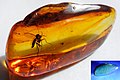 Baltic Amber.jpg 3,057 × 2,039; 1.2 MB
-
 Banner of Laboratory of Fluorescent Methods, National Laboratory Astana.jpg 5,000 × 1,971; 5.09 MB
-
 Blacklight-bodypaint.jpg 3,744 × 5,616; 6.22 MB
-
 C expcondition.png 315 × 140; 23 KB
-
 C fluorescence.png 257 × 257; 3 KB
-
 Calcite fluorescence.jpg 4,004 × 2,000; 4.57 MB
-
 Cb001.JPG 800 × 600; 145 KB
-
 Cell-universe.jpg 1,398 × 1,045; 51 KB
-
 Chinin Absorption Emission Spektrum.png 3,008 × 1,863; 233 KB
-
 Chinin Absorption Emission Spektrum.svg 1,052 × 744; 121 KB
-
 ClPhCz monokristall.jpg 1,575 × 1,169; 195 KB
-
 Cocktail straws.jpg 3,456 × 2,304; 4.14 MB
-
 Colonies de legionelles.jpg 700 × 694; 62 KB
-
 Colored balls - in a shop - Japan - Dec 2014.jpg 1,936 × 2,592; 738 KB
-
 Colored balls - zoom in - Japan - July 13 2015.jpg 1,387 × 1,140; 583 KB
-

-
 Connexion of neuves.jpg 1,376 × 1,032; 126 KB
-
 Correlative Microscopy with ZEISS Shuttle&Find - MERLIN and LSM 800 (16292616486).jpg 5,156 × 2,362; 976 KB
-
 Curcumin fluorescence.jpg 2,965 × 2,648; 513 KB
-
 Custom Suzuki GSXR (3796043588).jpg 960 × 720; 744 KB
-
 Cyanine J-aggregates formation.jpg 2,263 × 2,440; 541 KB
-
 DClPhCz kristall.jpg 2,048 × 1,536; 265 KB
-
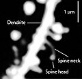 Dendritic spines.jpg 200 × 194; 25 KB
-
 Development of the retina.tif 6,560 × 1,312; 24.62 MB
-
 Diagrama de Jablonski.png 960 × 720; 55 KB
-
 Diagramme de Jablonski.png 1,353 × 1,013; 23 KB
-
 Diagramme partiel de Jablonski.png 637 × 788; 55 KB
-
 Dichlorofluoresceininnormallight.jpg 2,193 × 1,000; 303 KB
-
 DichlorofluoresceininUV.jpg 1,638 × 819; 242 KB
-
 Diindol.jpg 1,080 × 1,080; 146 KB
-
 Dna fluorescence.jpg 353 × 562; 132 KB
-
 DNA-conjugated gold nanoparticles +FAM DNA.png 439 × 294; 8 KB
-
 DNA-conjugated gold nanoparticles!.png 651 × 506; 25 KB
-
 DNA-conjugated gold nanoparticles.png 439 × 281; 6 KB
-
 DNA-conjugated nanoparticles +FAM DNA.png 716 × 554; 35 KB
-
 Dorsal Root Galglia Neurons.tif 6,144 × 1,920; 8.55 MB
-
 Droplet Fluorescence on Microwell Plate.jpg 320 × 105; 24 KB
-
 Drosophila wound healing.jpg 2,030 × 3,041; 860 KB
-
 DSC05968 k.jpg 2,762 × 2,511; 2.97 MB
-
 EB1911 - Fluorescence - Fig 1.jpg 209 × 230; 12 KB
-
 EB1911 - Fluorescence - Fig 2.jpg 166 × 177; 10 KB
-
 EB1911 - Fluorescence - Fig 3. Spectrum of Chlorophyll.jpg 339 × 197; 8 KB
-
 EB1911 - Fluorescence - Fig 4. Spectrum of Aesculinl.jpg 332 × 197; 15 KB
-
 EFluor Nanocrystal Vials.jpg 1,024 × 494; 119 KB
-
 Engin Umut Akkaya - BODIPY.JPG 2,000 × 3,008; 2.66 MB
-
 Engin Umut Akkaya - Reaction mechanism.JPG 2,000 × 2,921; 3.97 MB
-
 Epifluorescence microscopy of Elodea canadensis leaf hairs.jpg 4,148 × 2,765; 4.01 MB
-
 Epifluorescence microscopy of Elodea canadensis.jpg 5,184 × 3,456; 4.54 MB
-
 Erbium(III) chloride in fluorescent light.jpg 458 × 157; 13 KB
-
 EsfinjeFluor.jpg 3,240 × 4,320; 1.18 MB
-
 Espectro de excitación y de emisión de FITC.jpg 572 × 219; 31 KB
-
 Europium (III) Hydroxide under UV light.jpg 3,024 × 4,032; 894 KB
-
 FCS trace korrelation.png 800 × 1,157; 120 KB
-
 Fig1.gif 642 × 544; 7 KB
-
 FIRST measurement of SF6 and NH3.jpg 867 × 341; 18 KB
-
 Flames in bottle.jpg 750 × 1,000; 108 KB
-
 FlAVATAR carnations.jpg 5,472 × 3,648; 6.11 MB
-
 Flourescent nails (3746325122).jpg 3,264 × 2,448; 2.78 MB
-
 Fluo-phosopho.jpg 451 × 321; 12 KB
-
 Fluodual.jpg 328 × 215; 8 KB
-
 Fluorescence - phosphorescence, diagram.svg 1,119 × 785; 15 KB
-
 Fluorescence 1.jpg 2,724 × 3,092; 3.21 MB
-
 Fluorescence 4-methylumbelliferonu.jpg 719 × 1,280; 41 KB
-
 Fluorescence Dynamics and Photomanipulation (10690270154).jpg 5,903 × 8,259; 3.11 MB
-
 Fluorescence from Fluorescent Proteins.jpg 3,174 × 1,981; 804 KB
-
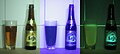
 Fluorescence in beer @ 450nm illumination.jpg 1,951 × 1,357; 467 KB
-
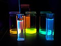
 Fluorescence in flasks.jpg 1,280 × 960; 112 KB
-
 Fluorescence in glow sticks that are yet to be activated.JPG 4,912 × 3,264; 2.31 MB
-
 Fluorescence in rhodamine B.jpg 2,400 × 1,600; 824 KB
-
 Fluorescence of Aesculin.JPG 2,272 × 1,704; 2.7 MB
-
 Fluorescence of Anthracene under UV light.jpg 2,822 × 2,845; 1.78 MB
-
 Fluorescence of chlorophyll under UV light.jpg 5,128 × 6,856; 15.8 MB
-
 Fluorescence of porphyrine.jpg 5,504 × 3,096; 3.67 MB
-
 Fluorescence of Terpyridine derivative.jpg 2,560 × 1,536; 1.13 MB
-
 Fluorescence off.jpg 3,150 × 1,717; 1.49 MB
-
 Fluorescence on a glass slide.jpg 743 × 917; 134 KB
-
 Fluorescence on.jpg 3,150 × 1,716; 1.44 MB
-
 Fluorescence resonance energy transfer.jpg 709 × 547; 115 KB
-
 Fluorescence.JPG 2,724 × 3,092; 2.92 MB
-
 Fluorescence.jpg 622 × 1,348; 173 KB
-
 FluorescenceMicroscopeSample HerringSpermSYBRGreen.jpg 2,633 × 1,800; 300 KB
-
 Fluorescencja białek.jpg 960 × 720; 73 KB
-
 Fluorescense Handful of light.jpg 3,308 × 2,482; 2.43 MB
-
 Fluorescense mix.JPG 2,724 × 3,092; 3.04 MB
-
 Fluorescense of Eysenhardtia polystachya's aqueous solution I.jpg 2,585 × 2,586; 744 KB
-
 Fluorescense of Eysenhardtia polystachya's aqueous solution II.jpg 2,515 × 3,354; 923 KB
-
 Fluorescent banana spots (4023036941).jpg 2,109 × 1,569; 541 KB
-
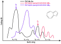
 Fluorescent Black-Light spectrum with peaks labelled-ru.svg 786 × 464; 35 KB
-
 Fluorescent Black-Light spectrum with peaks labelled.gif 800 × 515; 13 KB
-
 Fluorescent C. elangs D. melanogaster S. pombe.jpg 2,100 × 1,500; 560 KB
-
 Fluorescent Dyes and Proteins (10690288626).jpg 5,903 × 8,259; 2.02 MB
-
 Fluorescent leg warmers (3497879747).jpg 3,648 × 2,736; 2.21 MB
-
 Fluorescent Rocks.jpg 5,568 × 3,712; 1.76 MB
-
 Fluorescent Uranium Depression Glass.jpg 1,050 × 1,626; 805 KB
-
 Fluoresence.png 1,536 × 2,048; 1.83 MB
-
 Fluorestsents anisotroopia mõõtmine.jpg 336 × 278; 18 KB
-
 Fluoreszenz in Ethanol unter einer Quecksilberdampflampe - crop.jpg 262 × 255; 78 KB
-
 Fluoreszenz in Ethanol unter einer Quecksilberdampflampe.png 290 × 578; 356 KB
-
 Fluoreszenzlicht Plexiglas.png 491 × 453; 15 KB
-
 Fluoreszenzquenching von Chinin Spektrum.png 3,162 × 1,778; 93 KB
-
 Fluoreszenzquenching von Chinin Spektrum.svg 1,052 × 744; 359 KB
-
 Fluoreszierende Kunststoffplatte.jpg 1,584 × 1,564; 797 KB
-
 Fluoreszierende Stempeltinte.jpg 2,164 × 1,300; 621 KB
-
 Fluorexcitation.png 848 × 667; 18 KB
-
 Fluorimeter3D.jpg 600 × 423; 44 KB
-
 Fluorite fluorescence.jpg 4,004 × 2,000; 4.07 MB
-
 Fluorometro.JPG 360 × 192; 6 KB
-
 Fluorophore.png 619 × 482; 7 KB
-
 Fluorophors collection de.svg 826 × 336; 87 KB
-
 FP-Abb.png 775 × 1,230; 98 KB
-
 FPbeachTsien.jpg 830 × 810; 175 KB
-
 FRET Jablonski diagram.svg 733 × 715; 208 KB
-
 Galaxie.005.A.tif 993 × 757; 465 KB
-
 Galaxie.006.tif 692 × 685; 286 KB
-
 Galaxie.007.tif 1,017 × 896; 157 KB
-
 GFPmouse.jpg 4,288 × 3,216; 2.82 MB
-
 Glass fluorescence induced by 365nm light.JPG 4,928 × 3,264; 5.96 MB
-
 Glow in the Dark Bodypaint (8580024160).jpg 1,000 × 1,603; 1.05 MB
-
 Glowing Rocks.jpg 2,100 × 1,538; 1.35 MB
-
 Gold nanoparticles +FAM DNA!.png 580 × 392; 24 KB
-
 Gold nanoparticles +FAM DNA.png 439 × 309; 6 KB
-
 GreenBeamMeUp.jpg 1,024 × 575; 83 KB
-
 Greensub.jpg 1,536 × 2,048; 265 KB
-
 Gypsum fluorescence.jpg 3,456 × 5,184; 5.68 MB
-
 Hackmanite sous UVL.JPG 4,288 × 2,848; 3.73 MB
-
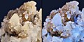
 Hackmanite, winchite sous UVL 1.JPG 4,288 × 2,848; 5.6 MB
-
 Hackmanite, winchite sous UVL 2.JPG 4,288 × 2,848; 3.69 MB
-
 High res fluorescence.jpg 514 × 320; 67 KB
-
 Highlighter in (green).jpg 1,600 × 900; 57 KB
-
 Highlighter inc (yellow).jpg 4,128 × 2,322; 1.29 MB
-
 HINA fluorophore.jpg 3,167 × 1,722; 4.71 MB
-
 Hulk green.jpg 1,504 × 1,000; 187 KB
-
 Hypholoma-fasciculare-Alan-Rockefeller-inaturalist-14376439.jpg 2,048 × 1,367; 1.06 MB
-
 Ibanez RG maple fretboard with Fluorescent Yellow in the case.jpg 800 × 600; 69 KB
-
 Imaging Life with Fluorescent Proteins (10690274384).jpg 5,531 × 6,876; 3.32 MB
-
 Jablonski Diagram of Fluorescence Only-ar.svg 648 × 865; 6 KB
-
 Jablonski Diagram of Fluorescence Only-de.png 576 × 861; 29 KB
-
 Jablonski Diagram of Fluorescence Only-en.svg 648 × 865; 2 KB
-
 Jablonski Diagram of Fluorescence Only-ru.svg 648 × 865; 2 KB
-
 Jablonski Diagram of Fluorescence Only.png 621 × 939; 22 KB
-
 Jablonski Diagram of Fluorescence und T1o.png 1,060 × 1,120; 62 KB
-
 Jablonski diagram rus.png 500 × 379; 26 KB
-
 JupilerFluo.jpg 3,330 × 1,304; 985 KB
-
 Kasha rule.svg 839 × 595; 19 KB
-
 Kasha-s-rule.png 378 × 378; 7 KB
-
 Kautsky effect.PNG 485 × 267; 8 KB
-
 Laser Fluorescence Cutaway Imaging Setup.jpg 2,048 × 1,536; 1.09 MB
-
 Laser Fluorescence Cutaway of Pentax Lens - Stretched.jpg 2,858 × 1,458; 1.3 MB
-
 Laser Fluorescence Cutaway of Pentax Lens.jpg 2,715 × 2,489; 3.14 MB
-
 Laser Fluorescence Imaging Setup.jpg 2,048 × 1,536; 1.15 MB
-
 Lazurite et afghanite sous UV (Sar-e-Sang, Koksha Valley, Badakshan - Afghanistan).JPG 3,563 × 2,683; 9.96 MB
-
 LBC Cover.jpg 600 × 800; 112 KB
-
 LeffeBlondeFluo.jpg 1,944 × 869; 384 KB
-
 LensFilter-001.jpg 2,400 × 1,800; 541 KB
-
 Les chélates et cryptates de terre rare..png 1,353 × 1,663; 36 KB
-
 LidarInelastique.jpg 1,000 × 603; 146 KB
-
 Light Demonstration.gif 600 × 338; 11.51 MB
-
 LIILIII-GIN-cells-show-typical-characteristics-of-Martinotti-cells.jpg 788 × 1,406; 311 KB
-
 Lucigenin.svg 750 × 1,125; 35 KB
-
 Lucigenin16-big.jpg 1,151 × 768; 148 KB
-
 Lucigenin17-big.jpg 1,151 × 768; 231 KB
-
 Lucygenina.png 154 × 152; 5 KB
-
 Luminous reaction.jpg 2,048 × 1,536; 49 KB
-
 Luray Caverns Gift Shop (8041013228) (2).jpg 4,592 × 2,576; 3.44 MB
-
 MAPbBr3 Nanocrystals Under UV.jpg 1,536 × 2,048; 197 KB
-

-
 Matlaline structure.svg 695 × 615; 22 KB
-
 Microphotograph of connexin 43 distribution in the rat myocardium.jpg 965 × 606; 631 KB
-
 MicroscopesOverview nl.jpg 1,322 × 900; 108 KB
-
 MicroscopesOverview.jpg 905 × 651; 128 KB
-
 NdCl3 fluolt.jpg 578 × 280; 23 KB
-
 NdCl3color.jpg 286 × 323; 25 KB
-
 Neuronal calcium.gif 513 × 511; 5.96 MB


.jpg)

.jpg)
.jpg)
.jpg)


.jpg)

.jpg)

.jpg)


_Hydroxide_under_UV_light.jpg)


.jpg)


.jpg)


.jpg)


.jpg)
.jpg)


.jpg)


.jpg)
.jpg)


.jpg)


.JPG)


_(2).jpg)


_chloride_in_fluorescent_light.jpg)