File:709 2017 1147 Fig6l.jpg
Jump to navigation
Jump to search

Size of this preview: 447 × 599 pixels. Other resolutions: 179 × 240 pixels | 358 × 480 pixels | 734 × 984 pixels.
Original file (734 × 984 pixels, file size: 72 KB, MIME type: image/jpeg)
Captions
Captions
Add a one-line explanation of what this file represents
Summary
[edit]| Description |
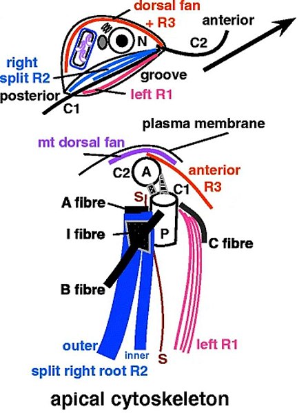
English: Cytoskeletal innovations during loukozoa origins. Ancestral condition in loukozoa summarized as represented by Malawimonas. Upper shows the whole cell seen from the right with the feeding groove tilted obliquely to show left and right mt roots (R1, R2) that support feeding groove rims and floor. Younger anterior cilium (C2) with oar-like beat and older posterior cilium (C1) undulating from base to tip simultaneously propel the cell forward (arrow) and waft food into the groove for ingestion. Lower diagram views the cell apex from the ventral side (so the cell’s right is on the left) to show mt arrays (colour: mt bands R1–R3; plus a dorsal fan of diverging mts that support the cell’s dorsal surface) and associated fibrous supports (black: A–C, I). The orthogonal centrioles (anterior A, posterior P) are interconnected by asymmetric linkers and in loukozoa (left) a dorsal mt fan and anterior left mt band (R3) connect C2s to the apical dorsal plasma membrane. R3 is developmental precursor of R1. The ancestral loukozoan interposed novel alveoli between the plasma membrane and dorsal fan, which split into a right bypassing mt band (BB) and numerous single, diverging subpellicular mts attached to alveolar inner faces.
The text argues that developmentally and evolutionarily the singlet root (S, brown) is a specialised R2 subcomponent, not a third posterior root as traditionally assumed. Dorsal fan and apical mts are actually longitudinal (as shown for BB only); the purple line symbolises a cross section of their mt arrays. |
| Date | |
| Source | Fig. 6 (left) at https://link.springer.com/article/10.1007/s00709-017-1147-3 Kingdom Chromista and its eight phyla: a new synthesis emphasising periplastid protein targeting, cytoskeletal and periplastid evolution, and ancient divergences. In: Protoplasma 255, pages 297–357,doi:10.1007/s00709-017-1147-3 |
| Author | Thomas Cavalier-Smith (caption slightly moved, digitylly enhanced) |
| Other versions |

Licensing
[edit]
This file is licensed under the Creative Commons Attribution-Share Alike 4.0 International license.
- You are free:
- to share – to copy, distribute and transmit the work
- to remix – to adapt the work
- Under the following conditions:
- attribution – You must give appropriate credit, provide a link to the license, and indicate if changes were made. You may do so in any reasonable manner, but not in any way that suggests the licensor endorses you or your use.
- share alike – If you remix, transform, or build upon the material, you must distribute your contributions under the same or compatible license as the original.
File history
Click on a date/time to view the file as it appeared at that time.
| Date/Time | Thumbnail | Dimensions | User | Comment | |
|---|---|---|---|---|---|
| current | 14:33, 8 August 2022 | | 734 × 984 (72 KB) | Ernsts (talk | contribs) | Uploaded a work by Thomas Cavalier-Smith from Fig. 3+6 at https://link.springer.com/article/10.1007/s00709-017-1147-3 Kingdom Chromista and its eight phyla: a new synthesis emphasising periplastid protein targeting, cytoskeletal and periplastid evolution, and ancient divergences. In: Protoplasma 255, doi:10.1007/s00709-017-1147-3 with UploadWizard |
You cannot overwrite this file.
File usage on Commons
The following page uses this file:
File usage on other wikis
The following other wikis use this file:
- Usage on de.wikipedia.org
- Usage on en.wikipedia.org
- Usage on hu.wikipedia.org