File:Aeolidiella stephanieae 12.png
Jump to navigation
Jump to search

Size of this preview: 625 × 600 pixels. Other resolutions: 250 × 240 pixels | 500 × 480 pixels | 672 × 645 pixels.
Original file (672 × 645 pixels, file size: 146 KB, MIME type: image/png)
Captions
Captions
Add a one-line explanation of what this file represents

|
This biology image could be re-created using vector graphics as an SVG file. This has several advantages; see Commons:Media for cleanup for more information. If an SVG form of this image is available, please upload it and afterwards replace this template with
{{vector version available|new image name}}.
It is recommended to name the SVG file “Aeolidiella stephanieae 12.svg”—then the template Vector version available (or Vva) does not need the new image name parameter. |
| Description |
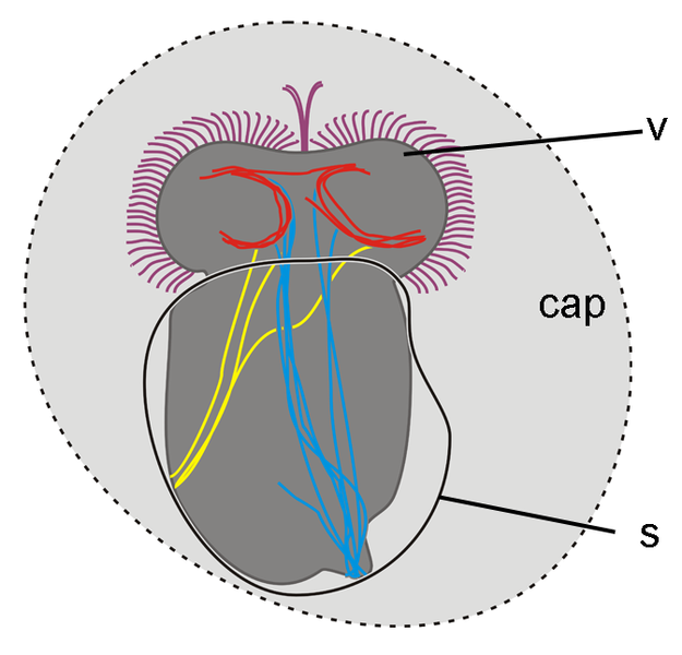
English: Drawing of an dorsal view of an early veliger stage larva (5% of development) of Berghia stephanieae (Valdés, 2005) (synonym: Aeolidiella stephanieae) showing first fibres of the larval retractor muscle (cyan), the accessory retractor muscle (yellow) and the velar ring muscles (red)
|
| Date | |
| Source | Kristof A. & Klussmann-Kolb A. (22 January 2010). "Neuromuscular development of Aeolidiella stephanieae Valdéz, 2005 (Mollusca, Gastropoda, Nudibranchia)". Frontiers in Zoology 7: 5. doi:10.1186/1742-9994-7-5. |
| Author | Alen Kristof & Annette Klussmann-Kolb |
| Other versions |
|

This file is licensed under the Creative Commons Attribution 2.0 Generic license.
- You are free:
- to share – to copy, distribute and transmit the work
- to remix – to adapt the work
- Under the following conditions:
- attribution – You must give appropriate credit, provide a link to the license, and indicate if changes were made. You may do so in any reasonable manner, but not in any way that suggests the licensor endorses you or your use.
File history
Click on a date/time to view the file as it appeared at that time.
| Date/Time | Thumbnail | Dimensions | User | Comment | |
|---|---|---|---|---|---|
| current | 00:29, 25 February 2010 | | 672 × 645 (146 KB) | Snek01 (talk | contribs) | {{Information |Description={{en|1=Drawing of an dorsal view of an early veliger stage larva (5% of development) of ''Aeolidiella stephanieae'' showing first fibres of the larval retractor muscle (cyan), the accessory retractor muscle (yellow) and the vela |
You cannot overwrite this file.
File usage on Commons
The following page uses this file:
File usage on other wikis
The following other wikis use this file:
- Usage on en.wikipedia.org